Copyright
©2006 Baishideng Publishing Group Co.
World J Gastroenterol. Feb 28, 2006; 12(8): 1165-1174
Published online Feb 28, 2006. doi: 10.3748/wjg.v12.i8.1165
Published online Feb 28, 2006. doi: 10.3748/wjg.v12.i8.1165
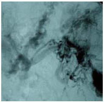
Figure 1 Conventional splenoportography showing a case of portal cavernomatous transformation with portosystemic collaterals, and extensive esophageal-gastric varicose veins.
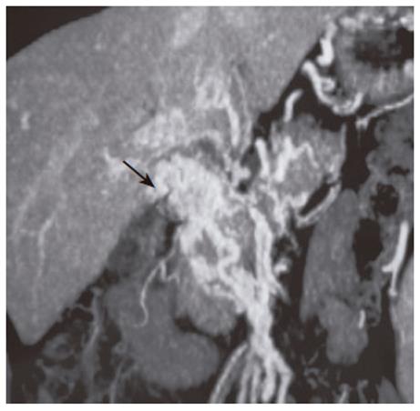
Figure 2 CT-angiography of portal system.
Arrow shows the portal cavernomatous transformation with portosystemic collaterals.
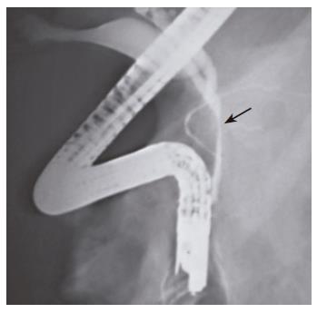
Figure 3 ERCP of a patient with portal cavernomatous transformation.
Arrow shows the site of depression in the main bile duct.
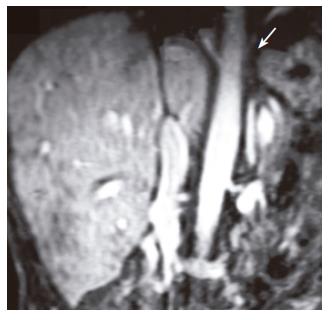
Figure 4 An MRI venography of hepatic veins and inferior vena cava.
Arrow shows the site of obstruction in vena cava and unvisible hepatic veins.
- Citation: Bayraktar Y, Harmanci O. Etiology and consequences of thrombosis in abdominal vessels. World J Gastroenterol 2006; 12(8): 1165-1174
- URL: https://www.wjgnet.com/1007-9327/full/v12/i8/1165.htm
- DOI: https://dx.doi.org/10.3748/wjg.v12.i8.1165


