Copyright
©2006 Baishideng Publishing Group Co.
World J Gastroenterol. Feb 21, 2006; 12(7): 1149-1152
Published online Feb 21, 2006. doi: 10.3748/wjg.v12.i7.1149
Published online Feb 21, 2006. doi: 10.3748/wjg.v12.i7.1149
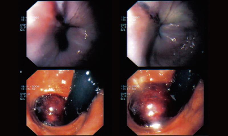
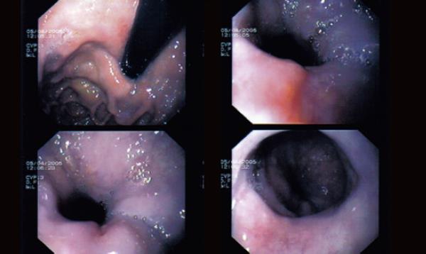
Figure 1 upper GI endoscopy A big hemorrhagic polypoid lesion as a haematoma in the hiatus hernia at upper GI endoscopy
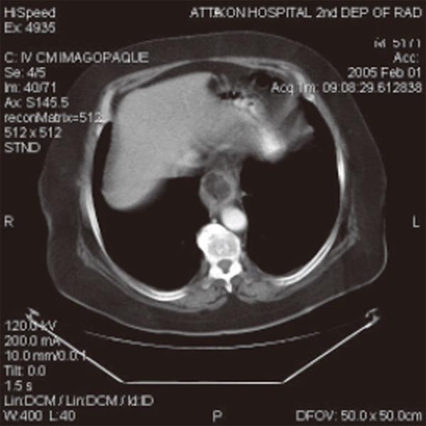
Figure 2 Intraluminal mass with a lipoma-like density in the lower esophagus.
Figure 3 The lesion is covered by yellowish exudates at the second upper GI endoscopy.
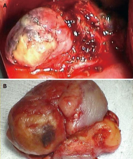
Figure 4 Intraoperative view of the esophageal liposarcoma (A) and the specimen (B) (5 cm x 3.
5 cm x 2 cm).
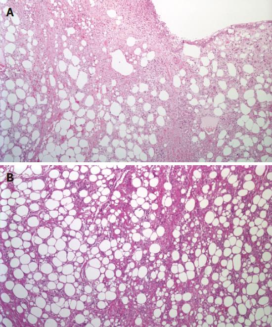
Figure 5 Neoplasm infiltrating smooth muscle layer of esophagus and ulcerating surface epithelium (A),HE 100x; and neoplasm consisting of differently-sized lipocytic element and multi-vacuolated lipoblasts (B), HE 400x.
A small amount of sclerotic stroma was focally identified with the presence of atypical stromal cells.
Figure 6 No evidence of tumor recurrence in gastrointestinal endoscopy after 6 mo of surgery.
- Citation: Liakakos TD, Troupis TG, Tzathas C, Spirou K, Nikolaou I, Ladas S, Karatzas GM. Primary liposarcoma of esophagus: A case report. World J Gastroenterol 2006; 12(7): 1149-1152
- URL: https://www.wjgnet.com/1007-9327/full/v12/i7/1149.htm
- DOI: https://dx.doi.org/10.3748/wjg.v12.i7.1149


