Copyright
©2006 Baishideng Publishing Group Co.
World J Gastroenterol. Dec 21, 2006; 12(47): 7598-7603
Published online Dec 21, 2006. doi: 10.3748/wjg.v12.i47.7598
Published online Dec 21, 2006. doi: 10.3748/wjg.v12.i47.7598
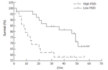
Figure 1 Kaplan-Meier survival curve correlating disease specific survival with high microvessel density (MVD) or low MVD.
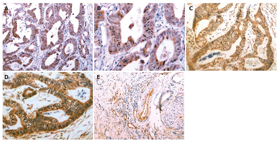
Figure 2 Immunohistochemical stainings of COX-2 (A, B), VEGF (C, D) and microvessels (E) in tissue sections obtained from gastric adenocarcinoma.
COX-2 was mainly expressed in the cytoplasm of cancer cells (brown staining; A × 100, B × 200). VEGF expression was restricted to the cytoplasm of cancer cells (brown staining; C × 100, D × 200). Microvessels were detected in gastric cancer tissues by immunostaining for factor VIII-related antigen (E × 100).
- Citation: Zhao HC, Qin R, Chen XX, Sheng X, Wu JF, Wang DB, Chen GH. Microvessel density is a prognostic marker of human gastric cancer. World J Gastroenterol 2006; 12(47): 7598-7603
- URL: https://www.wjgnet.com/1007-9327/full/v12/i47/7598.htm
- DOI: https://dx.doi.org/10.3748/wjg.v12.i47.7598


