Copyright
©2006 Baishideng Publishing Group Co.
World J Gastroenterol. Dec 14, 2006; 12(46): 7451-7459
Published online Dec 14, 2006. doi: 10.3748/wjg.v12.i46.7451
Published online Dec 14, 2006. doi: 10.3748/wjg.v12.i46.7451
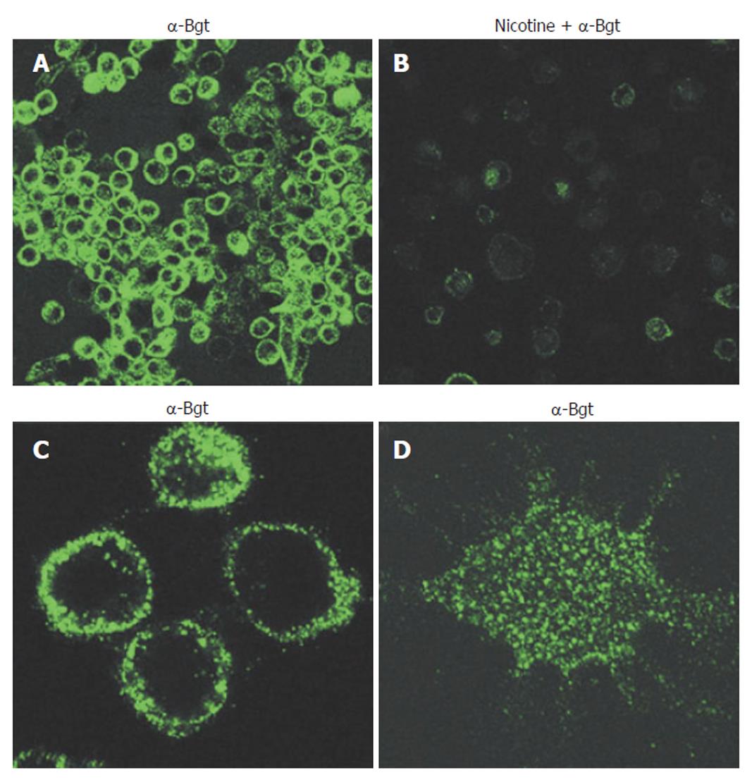
Figure 1 α-Bungarotoxin-binding nicotinic receptors are clustered on the surface of macrophages.
Primary human macrophages were stained with fluorescein isothiocyanate (FITC)-labelled α-bungarotoxin (1.5 μg /mL) and viewed by fluorescent confocal microscopy. A: Cells were stained with α-bungarotoxin alone; B: Nicotine was added to a final concentration of 500 μmol before addition of α-bungarotoxin. C, D: Higher magnification reveals receptor clusters. C: Focus planes are on the inside layers close to the middle (three lower cells) or close to the surface (upper cell) of cells; D: Focus plane is on the surface of the cell. Magnifications: A, B, x 50; C, x 200; D, x 450.
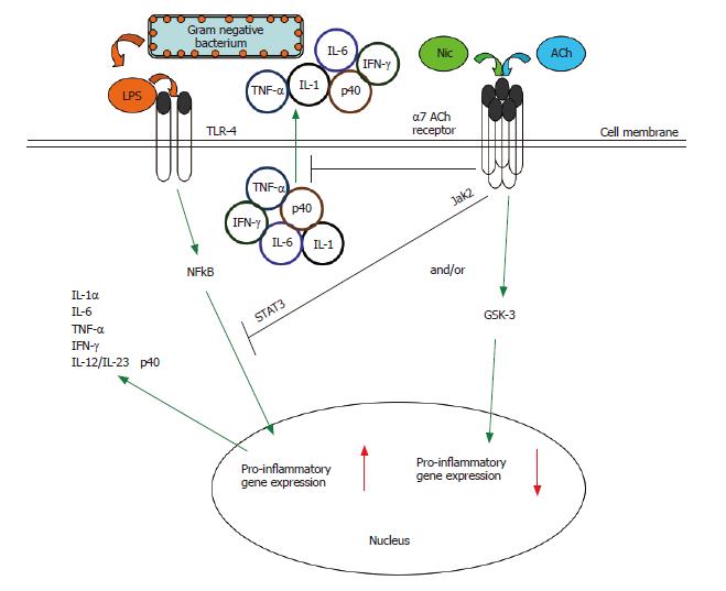
Figure 2 The cholinergic anti-inflammatory pathway.
Multiple inflammatory stimuli activate the NFκB system and lead to the release of pro-inflammatory cytokines from innate immune cells. For example, interaction of bacterial LPS with Toll-like receptors (TLRs) on the monocyte surface induces a pro-inflammatory response characterized by the production and release of several key pro-inflammatory cytokines[88]. The α7 nAChR-dependent cholinergic anti-inflammatory pathway, triggered endogenously by acetylcholine or exogenously by nicotine, can suppress the production of several pro-inflammatory cytokines in activated monocytic cells (see Figure 4)[21,22,32,39,45]. Such nicotine-mediated suppression of TNF in vivo protects mice from endotoxic shock[32,46]. The cholinergic anti-inflammatory pathway acts at both the transcriptional and post-translational levels. Engagement of the α7 nAChR results in the rapid suppression of the release of pre-formed pro-inflammatory cytokines[32,39,45-47]. Engagement of the α7 nAChR also results in inactivation of the NFκB system, preventing the upregulation of pro-inflammatory gene activity[32]. There is a need to further explore the signaling within the cholinergic anti-inflammatory pathway. In macrophages, the nicotine-induced suppression of pro-inflammatory cytokine release involves recruitment of the tyrosine kinase Jak2 to the α7 nACh receptor, the subsequent phosphorylation of the transcription factor STAT3, and the activation of STAT3 and SOC3 signaling cascade[61]. We have shown the potential convergence of the nicotinic anti-inflammatory and an endogenous, GSK-3-dependent anti-inflammatory pathway[88] in monocytes (see Figure 5).
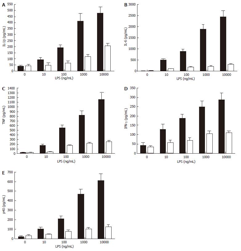
Figure 4 Nicotine inhibits the release of multiple cytokines (TNF, IFN-γ, IL-1β, IL-6, and the common IL-12/IL-23 subunit, p40) under the control of the NF-κB pathway.
Cells were pre-treated with nicotine (100 ng/mL) for 2 h then stimulated with purified LPS (0 to 1 x 10⁴ ng/mL) for 24 h. Cell-free supernatants were harvested by centrifugation and levels of pro-inflammatory cytokines were determined by ELISA. Data represents the mean (SD) of triplicate experiments.
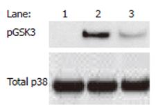
Figure 5 Nicotine-treated human monocytes exhibit augmented levels of phosphorylated (Ser9) GSK3-β in monocytes stimulated with the Gram negative bacterium, Porphyromonas gingivalis.
Monocytes were pre-treated for 2 h with 100 ng/mL of nicotine and stimulated for 60 min with P. gingivalis (MOI = 10). Western blot was performed using whole-cell lysates (20 μg) and probing for GSK3 using a phospho-specific GSK3-β (Ser9; denoted pGSK3) antibody. Blot was stripped and re-probed for total p38 to ensure equivalent loading. Lane 1: Nonstimulated; Lane 2: P. gingivalis + Nicotine; Lane 3: P. gingivalis. Data are representative of three experiments.
Figure 3 Periodontitis in a male smoker, age 55.
A: An anterior view of the mouth of a male smoker, age 55. The teeth have some staining and visible plaque. The gingivae are receded and some root surfaces are exposed. The gingivae in the upper jaw are relatively uninflamed and appear pink and fibrous in contrast to the red and swollen appearance in the lower anterior jaw; B: Radiographs of the lower anterior teeth. One tooth exfoliated a few months before. The remaining incisor teeth have advanced bone loss almost to the apices. Loss of these teeth is almost inevitable.
- Citation: Scott DA, Martin M. Exploitation of the nicotinic anti-inflammatory pathway for the treatment of epithelial inflammatory diseases. World J Gastroenterol 2006; 12(46): 7451-7459
- URL: https://www.wjgnet.com/1007-9327/full/v12/i46/7451.htm
- DOI: https://dx.doi.org/10.3748/wjg.v12.i46.7451


