Copyright
©2006 Baishideng Publishing Group Co.
World J Gastroenterol. Oct 7, 2006; 12(37): 5972-5977
Published online Oct 7, 2006. doi: 10.3748/wjg.v12.i37.5972
Published online Oct 7, 2006. doi: 10.3748/wjg.v12.i37.5972
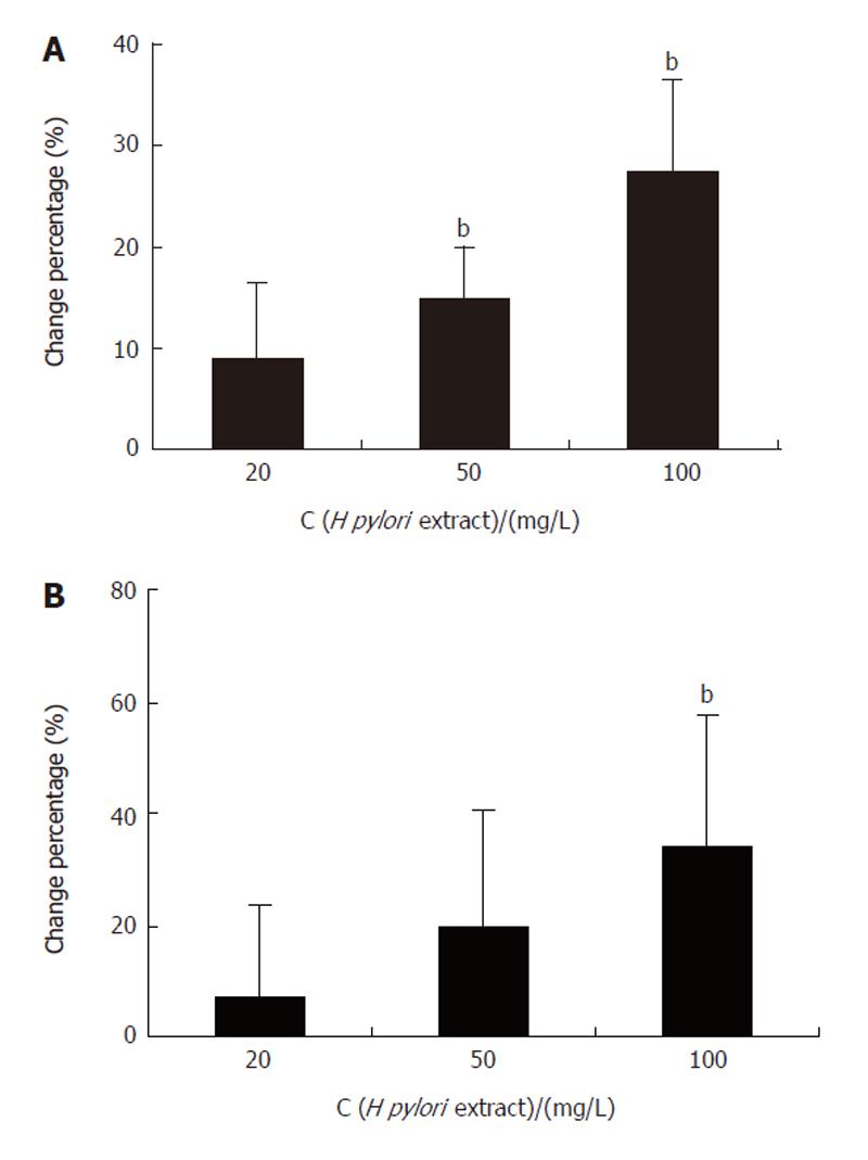
Figure 1 MTT assay (A) and 3H-TdR-incorporation test (B) showing H pylori extract- stimulated proliferation of BGC-823 cells.
bP < 0.01 vs control.
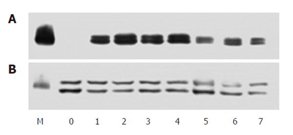
Figure 2 Western blotting showing phosphorylated ERK (A) and total ERK (B) in serum-starved BGC-823 cells incubated with H pylori extract.
M: Protein molecular marker; 0: Control; 1-7: Incubation with 50 mg/L H pylori extract for 20, 40, 60 min and 3, 6, 12, 24 h.
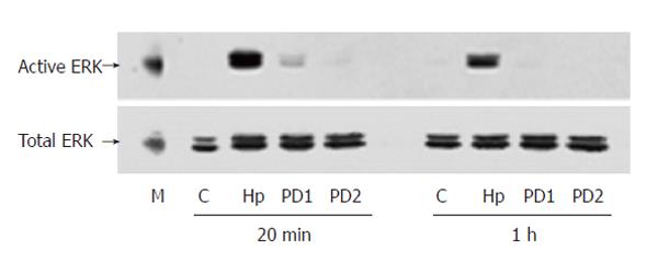
Figure 3 Western blotting showing PD98059-blocked stimulating effect of H pylori extract on ERK activation in BGC-823 cells.
M: Molecular marker; C: Control; Hp: 50 mg/L H pylori extract; PD1: 25 μmol/L PD98059 + 50 mg/L H pylori extract; PD2: 50 μmol/L PD98059 + 50 mg/L H pylori extract.
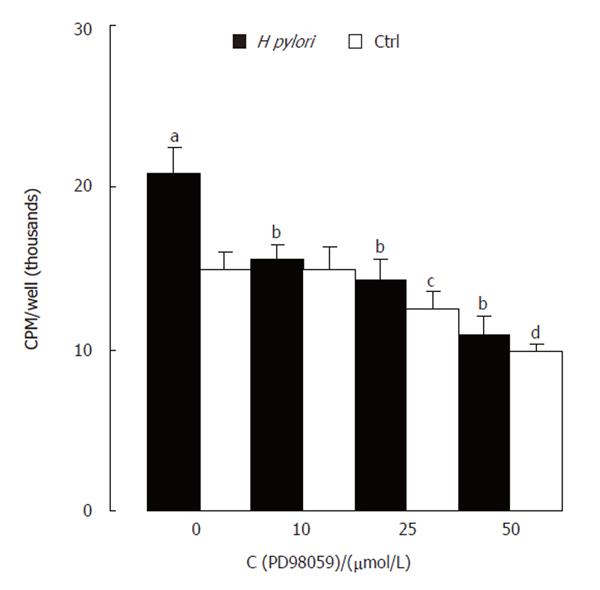
Figure 4 MTT assay showing PD98059-blocked proliferation-stimulating effect of H pylori extract.
aP < 0.05, cP < 0.05, dP < 0.01 vs control only; bP < 0.01 vs Hp only.
Figure 5 Western blotting showing genistein-prevented ERK activation by H pylori extract.
M: Molecular marker; C: Control; Hp: H pylori extract; Hp + G: Genistein and H pylori extract.
Figure 6 Western blotting showing H pylori extract-caused tyrosine pho-sphorylation of cell lysate of BGC-823.
M: Molecular marker; 1: control; 2-7: H pylori extract incubated for 20, 40 min and 1, 3, 6, 12 h.
Figure 7 Western blotting showing H pylori extract- increased expression of c-Fos.
1: Control; 2-8: H pylori extract incubated for 20, 40 min and 1, 3, 6, 12 and 24 h.
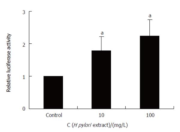
Figure 8 H pylori extract-stimulated SRE-dependent reporter gene expression.
aP < 0.05 vs control.
- Citation: Chen YC, Wang Y, Li JY, Xu WR, Zhang YL. H pylori stimulates proliferation of gastric cancer cells through activating mitogen-activated protein kinase cascade. World J Gastroenterol 2006; 12(37): 5972-5977
- URL: https://www.wjgnet.com/1007-9327/full/v12/i37/5972.htm
- DOI: https://dx.doi.org/10.3748/wjg.v12.i37.5972


