Copyright
©2006 Baishideng Publishing Group Co.
World J Gastroenterol. Sep 28, 2006; 12(36): 5793-5797
Published online Sep 28, 2006. doi: 10.3748/wjg.v12.i36.5793
Published online Sep 28, 2006. doi: 10.3748/wjg.v12.i36.5793
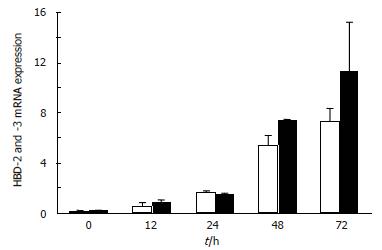
Figure 1 Time course of human β-defensin mRNA expression induced by H pylori in a gastric cancer cell line MKN45.
MKN45 cells (106) were seeded into dishes 60 mm in diameter and incubated for 12 h. Culture medium was replaced with 2 mL of fresh RPMI 1640 medium without FBS. The cells were incubated for 0 to 72 h with 100 μL of 108 CFU/mL H pylori. Human β-defensin mRNA expression was measured using TaqMan RT-PCR assay. hBD-2 (□), hBD-3 (■).
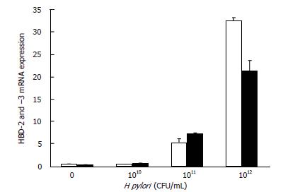
Figure 2 Induction of human β-defensin mRNA expression by various numbers of H pylori.
MKN45 cells (106) were seeded into dishes 60 mm in diameter and incubated for 12 h. Culture medium was replaced with 2 mL of fresh RPMI 1640 medium without FBS. Bacterial suspensions (100 μL; 0 to 109 CFU/mL in RPMI 1640 medium) were added to the dishes, and incubation was continued for 48 h. Human β-defensin mRNA expression was measured using TaqMan RT-PCR assay. hBD-2 (□), hBD-3 (■).
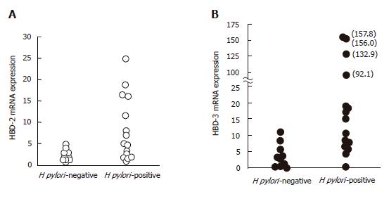
Figure 3 Human β-defensin mRNA expressions in gastric mucosal tissues from 15 H pylori-positive patients (○) and 10 H pylori-negative patients (●).
Human β-defensin mRNA expression was measured using TaqMan RT-PCR assay. A: hBD-2; B: hBD-3.
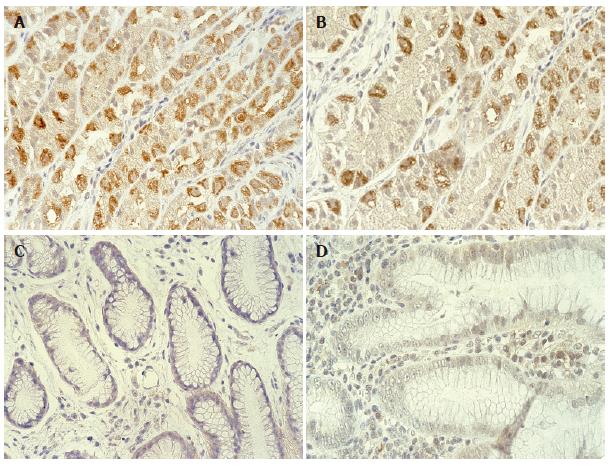
Figure 4 Human β-defensin protein expressions in gastric mucosal tissues with or without H pylori infection.
Tissues were stained with anti-hBD-2 antibody (A and C) or hBD-3 antibody (B and D). Case 1: A and B, gastric mucosa with H pylori -associated gastritis. Immunostaining was observed in gastric cancer cells in A and gastric epithelial cells in B. Case 2: C and D, gastric mucosa with gastritis but without H pylori infection. No staining was observed in C and D.
Figure 5 Antimicrobial effects of human β-defensin protein on H pylori (ATCC49504).
H pylori were cultured on HP agar for 4 d after a 1-h pre-incubation in the presence or absence of hBD-2 (A) and hBD-3 (B).
- Citation: Kawauchi K, Yagihashi A, Tsuji N, Uehara N, Furuya D, Kobayashi D, Watanabe N. Human β-defensin-3 induction in H pylori-infected gastric mucosal tissues. World J Gastroenterol 2006; 12(36): 5793-5797
- URL: https://www.wjgnet.com/1007-9327/full/v12/i36/5793.htm
- DOI: https://dx.doi.org/10.3748/wjg.v12.i36.5793


