Copyright
©2006 Baishideng Publishing Group Co.
World J Gastroenterol. Sep 21, 2006; 12(35): 5733-5734
Published online Sep 21, 2006. doi: 10.3748/wjg.v12.i35.5733
Published online Sep 21, 2006. doi: 10.3748/wjg.v12.i35.5733
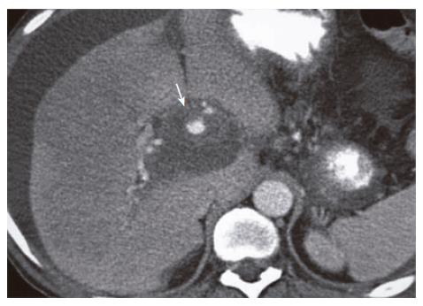
Figure 1 Contrast-enhanced CAT of the abdomen showing a 6 cm × 6 cm pseudoaneurysm of the proper hepatic artery.
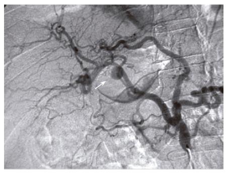
Figure 2 Angiogram showing an aneurysmal sac with a narrow neck originating from the inferior aspect of the distal portion of the proper hepatic artery.
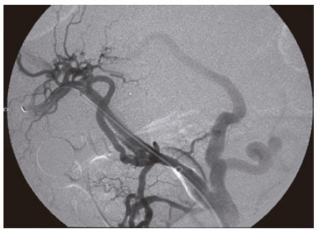
Figure 3 Repeat angiogram demonstrating complete exclusion of the aneurysm.
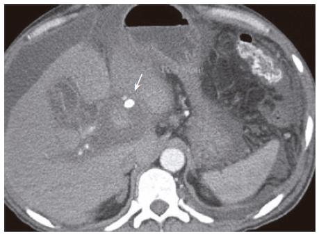
Figure 4 A repeat CAT scan of the abdomen after 24 h showing successful stenting.
- Citation: Singh CS, Giri K, Gupta R, Aladdin M, Sawhney H. Successful management of hepatic artery pseudoaneurysm complicating chronic pancreatitis by stenting. World J Gastroenterol 2006; 12(35): 5733-5734
- URL: https://www.wjgnet.com/1007-9327/full/v12/i35/5733.htm
- DOI: https://dx.doi.org/10.3748/wjg.v12.i35.5733


