Copyright
©2006 Baishideng Publishing Group Co.
World J Gastroenterol. Sep 21, 2006; 12(35): 5651-5657
Published online Sep 21, 2006. doi: 10.3748/wjg.v12.i35.5651
Published online Sep 21, 2006. doi: 10.3748/wjg.v12.i35.5651
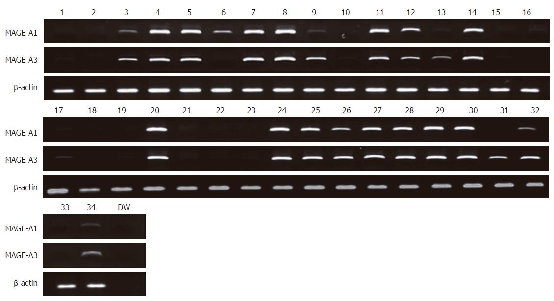
Figure 1 RT-PCR analysis of the MAGE-A1 and MAGE-A3 genes in 32 colorectal cancer cell lines.
β-actin was amplified as an internal control. The MAGE-A1 gene was significantly expressed in 19 colorectal cancer cell lines (Lanes, 3, 4, 5, 6, 7, 8, 9, 11, 12, 14, 20, 24, 25, 26, 27, 28, 29, 30 and 32). The MAGE-A3 gene was expressed in 21 colorectal cancer cell lines (Lanes, 3, 4, 5, 7, 8, 9, 11, 12, 13, 14, 17, 20, 24, 25, 26, 27, 28, 29, 30, 31 and 32). Lane numbers 1 to 34 show cell lines SNU-61, SNU-81, SNU-175, SNU-283, SNU-407, SNU-503, SNU-769A, SNU-769B, SNU-1033, SNU-1040, SNU-1047, SNU-1197, SNU-C1, SNU-C2A, SNU-C4, SNU-C5, Caco-2, COLO201, COLO205, COLO320, DLD1, HCT-8, HCT-15, HCT-116, HT-29, LOVO, LS174T, NCI-H716, SW403, SW480, SW1116, WiDr, SNU-1, and SNU-5 respectively.
Figure 2 Methylation analysis of the MAGE-A1 and MAGE-A3 genes in colorectal cancer cell lines.
The promoter region of the MAGE-A1 gene was unmethylated in 26 cell lines (SNU-61, SNU-175, SNU-283, SNU-503, SNU-769A, SNU-769B, SNU-1033, SNU-1197A, SNU-C1, SNU-C2A, SNU-C5, Caco2, COLO201, COLO205, COLO320, DLD-1, HCT-15, HCT-116, HT-29, Lovo, LS174T, NCI-H716, SW403, SW1116, and WiDR cell lines). Unmethylated MAGE-A3 DNA amplifications were found in 26 cell lines (SNU-61, SNU-175, SNU-407, SNU-503, SNU-769A, SNU-769B, SNU-1033, SNU-1047, SNU-1197A, SNU-C1, SNU-C2A, SNU-C4, Caco2, COLO201, COLO205, COLO320, DLD-1, HCT-15, HCT-116, HT-29, Lovo, LS174T, NCI-H716, SW403, SW1116, and WiDR cell lines). Lane M denotes product amplified by primers recognizing a methylated sequence and Lane U denotes the product amplified by primers recognizing an unmethylated sequence, respectively.
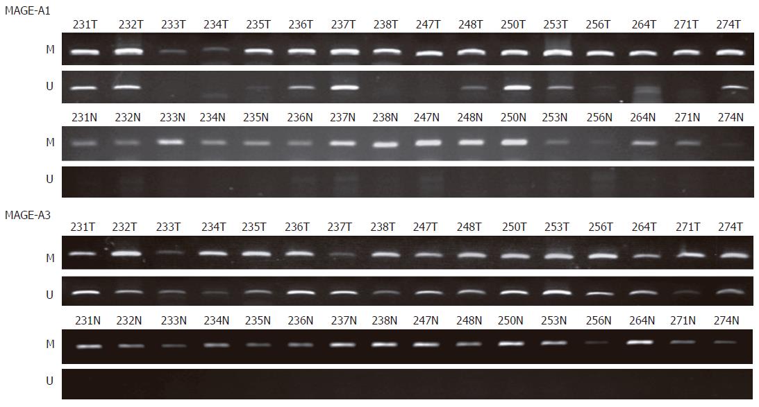
Figure 3 Methylation analysis of the MAGE-A1 and MAGE-A3 genes in colorectal cancer tissues and corresponding normal tissues.
Methylation-specific PCR product amplified by primers recognizing methylated and unmethylated sequence. The promoter region of the MAGE-A1 and MAGE-A3 genes was unmethylated in colorectal cancer tissues, not normal tissues. Numbers represent each colorectal tissue and T denotes colorectal tumor tissues and N denotes corresponding normal tissues.
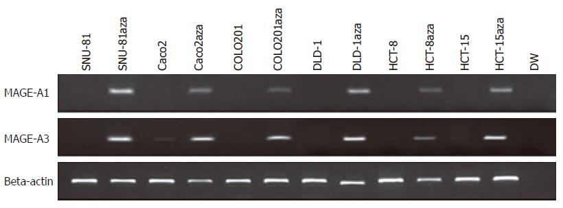
Figure 4 RT-PCR analysis after treatment with 5-aza-2’-deoxycytidine.
The MAGE-A1 and MAGE-A3 genes were reactivated.
-
Citation: Kim KH, Choi JS, Kim IJ, Ku JL, Park JG. Promoter hypomethylation and reactivation of
MAGE-A1 andMAGE-A3 genes in colorectal cancer cell lines and cancer tissues. World J Gastroenterol 2006; 12(35): 5651-5657 - URL: https://www.wjgnet.com/1007-9327/full/v12/i35/5651.htm
- DOI: https://dx.doi.org/10.3748/wjg.v12.i35.5651


