Copyright
©2006 Baishideng Publishing Group Co.
World J Gastroenterol. Sep 14, 2006; 12(34): 5429-5439
Published online Sep 14, 2006. doi: 10.3748/wjg.v12.i34.5429
Published online Sep 14, 2006. doi: 10.3748/wjg.v12.i34.5429
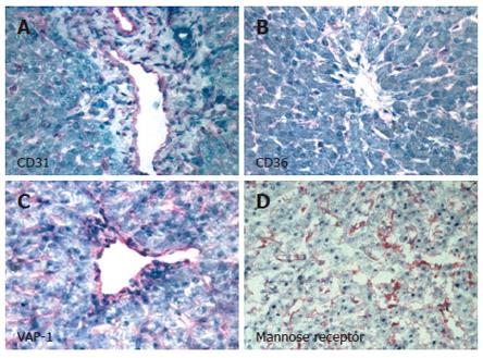
Figure 1 Human hepatic sinusoidal endothelial cells in situ express the classical endothelial phenotypic markers CD31 (A) and CD36 (B) as well as more recently identified markers such as VAP-1 (C) and Mannose receptor (D).
Images represent immunohistochemical staining of human liver sections using specific primary antibodies in an indirect immunoperoxidase protocol with haematoxylin counterstain. Positive staining of sinusoidal endothelial cells is indicated by pink pigmentation. CD31, CD36 and VAP-1 are observed on both portal vessel endothelial cells and sinusoidal endothelium, whilst the mannose receptor is localised on sinusoidal endothelial cells and Kupffer cells.
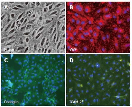
Figure 2 Isolated, cultured human hepatic sinusoidal endothelial cells exhibit classical morphology under phase contrast microscopy (A), and stain positively with antibody directed against endothelial phenotypic markers.
Images represent immunofluorescent staining of cultured cells using specific primary antibodies in an indirect fluorescent protocol with DAPI nuclear counterstain (blue). Positive staining for vWF (B) is visualised using a Texas Red-labelled secondary antibody, whilst expression of endoglin (CD105, C) and ICAM-2 (D) are visualised with a FITC-conjugated secondary antibody (green).
Figure 3 Cultured human hepatic sinusoidal endothelial cells stain positively with antibody directed against ‘non-classical endothelial’ phenotypic markers.
Images represent immunofluorescent staining of cultured cells using specific primary antibodies in an indirect fluorescent protocol. Expression of VAP-1 (A) and LYVE-1 (B) are visualised with a FITC-conjugated secondary antibody (green), whilst positive staining for L-SIGN (C) is visualised using a Texas Red-labelled secondary antibody.
- Citation: Lalor P, Lai W, Curbishley S, Shetty S, Adams D. Human hepatic sinusoidal endothelial cells can be distinguished by expression of phenotypic markers related to their specialised functions in vivo. World J Gastroenterol 2006; 12(34): 5429-5439
- URL: https://www.wjgnet.com/1007-9327/full/v12/i34/5429.htm
- DOI: https://dx.doi.org/10.3748/wjg.v12.i34.5429


