Copyright
©2006 Baishideng Publishing Group Co.
World J Gastroenterol. Sep 7, 2006; 12(33): 5404-5407
Published online Sep 7, 2006. doi: 10.3748/wjg.v12.i33.5404
Published online Sep 7, 2006. doi: 10.3748/wjg.v12.i33.5404
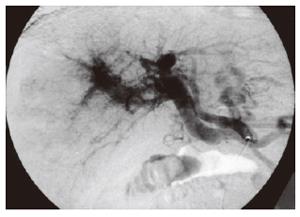
Figure 1 Celiac arteriography showing a marked arterioportal shunt with tumorous thread and streak sign and massive reverse flow to the main trunk of the portal vein.
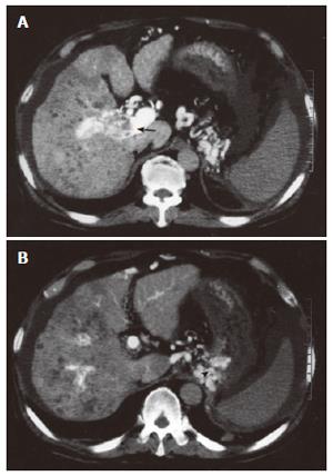
Figure 2 CT during proper hepatic arteriography showing tumor thrombi (arrow) in the right portal vein (A) and gastric varices (arrowhead) (B).
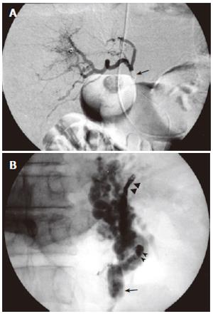
Figure 3 BRTO combined with temporary balloon occlusion of the hepatic artery.
A: Proper hepatic arteriography through the balloon catheter during balloon occlusion (arrow); B: Balloon-occluded retrograde venography (arrow) after embolization of the left inferior phrenic vein and the retroperitoneal vein with micro-spring coils (arrowheads) showing the stagnant gastrorenal shunt and gastric varices. A 5% ethanolamine oleate mixture (30 mL) was then injected during dual balloon occlusion.
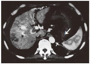
Figure 4 Contrast-enhanced CT showing thrombosed gastric varices (white arrow) on the following day.
- Citation: Nakai M, Sato M, Tanihata H, Sonomura T, Sahara S, Kawai N, Kimura M, Terada M. Ruptured high flow gastric varices with an intratumoral arterioportal shunt treated with balloon-occluded retrograde transvenous obliteration during temporary balloon occlusion of a hepatic artery. World J Gastroenterol 2006; 12(33): 5404-5407
- URL: https://www.wjgnet.com/1007-9327/full/v12/i33/5404.htm
- DOI: https://dx.doi.org/10.3748/wjg.v12.i33.5404


