Copyright
©2006 Baishideng Publishing Group Co.
World J Gastroenterol. Sep 7, 2006; 12(33): 5393-5395
Published online Sep 7, 2006. doi: 10.3748/wjg.v12.i33.5393
Published online Sep 7, 2006. doi: 10.3748/wjg.v12.i33.5393
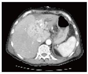
Figure 1 CT scan shows Klatskin tu-mor type III b with intrahepatic bile duct enlargement, cholesta-sis and atrophy of the left hepatic lobe.
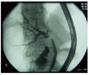
Figure 2 ERC dis-plays good filling of the right intrahepatic bile duct system whereas the left system could not be displayed due to tumorous stenosis in the distal part of the ductus hepaticus sinister reaching into the ductus hepaticus communis.
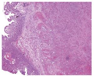
Figure 3 Histological examination of the resected colonic neoplasm reveal intra-mural metastasis of adenocarcinoma infiltrating the muscular and submucosal layers.
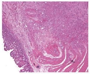
Figure 4 Histological examination of the resected colonic neoplasm reveals intramural metastasis of adenocarcinoma infiltrating the muscular and submucosal layers.
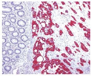
Figure 5 Immuno-histological staining shows adenocarcinoma of the non-intestinal klatskinoide type.
- Citation: Schmeding M, Neumann U, Neuhaus P. Colonic metastasis of Klatskin tumor: Case report and discussion of the current literature. World J Gastroenterol 2006; 12(33): 5393-5395
- URL: https://www.wjgnet.com/1007-9327/full/v12/i33/5393.htm
- DOI: https://dx.doi.org/10.3748/wjg.v12.i33.5393


