Copyright
©2006 Baishideng Publishing Group Co.
World J Gastroenterol. Aug 28, 2006; 12(32): 5168-5174
Published online Aug 28, 2006. doi: 10.3748/wjg.v12.i32.5168
Published online Aug 28, 2006. doi: 10.3748/wjg.v12.i32.5168
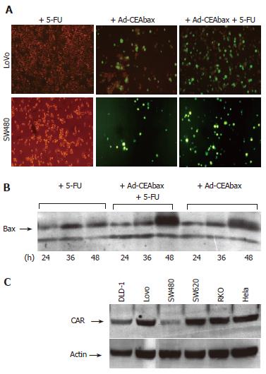
Figure 1 Effect of 5-FU on the adenoviral infection of colon carcinoma cell lines differing in CAR expression.
The drug was used at the dose of 2 μmol/L for the Lovo cells and of 20 μmol/L for the SW480 cells. The virus (Ad-CEAbax) was used at 1 MOI. The cells were treated with 5-FU alone, or infected with the virus in the absence and in the presence of 5-FU (A). The expression of bax with 5-FU and/or the virus was controlled by western blot after 24 h, 36 h and 48 h incubation (B). The expression level of CAR on the colon carcinoma cells line was confirmed by western blot analysis (the expression of actin is reported as a control for loading) (C).
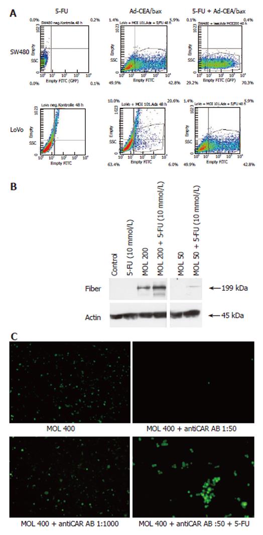
Figure 2 The number of GFP expression cells was measured by flow cytometry in SW480 and Lovo cells (A).
Treatment with 5-FU resulted in increased number of GFP expression cells and increased intensity of GFP expression in both cell lines. Intracellular adenoviral fiber protein was assessed by western blot analysis of SW480 cell lysates (B) after Ad-GFP treatment. In control cells and 5-FU treated cells no fiber protein could be detected. Low amounts of fiber protein could be seen in Ad-GFP treated cells dependent on Ad-GFP concentrations (MOI of 200 and 50). Additional 5-FU treatment increased fiber protein in whole cell lysates indicating enhanced adenoviral uptake. Function of CAR was blocked by anti CAR antibody at a concentration of 1:500 but 5-FU enhanced the number of GFP expressing cells even at anti CAR antibody concentrations of 1:50 (C).
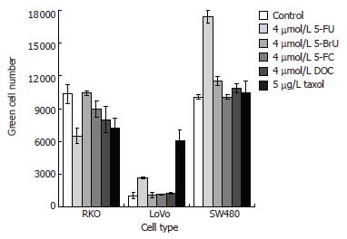
Figure 4 Other pyrimidine-based drugs do not show the positive effect of 5-FU on adenoviral infection.
The colon carcinoma cell lines were treated concurrently with the virus Ad-GFP and 5-FU, 5-BrU, 5-FC, DOC or taxol, each at the indicated concentration. The number of the infected green cells was counted after 40 h infection. The reported data are the average of three experiments and the error bars indicate the standard deviation values.
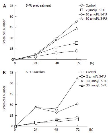
Figure 3 Dependence of adenoviral efficacy on the sequencing of 5-FU and virus administration to the SW480 cells.
Two h postincubation with the drug at the indicated concentrations, the drug was removed and the cells were infected with the adenoviral construct Ad-GFP (A). Alternatively, the cells were infected with the virus in combination with 5-FU (B). The number of green cells was counted after 24, 48 and 72 h infection. The reported data points are the average of three experiments and the standard deviation values were in the range of 10%-15% (the error bars were not reported for clarity).
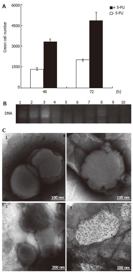
Figure 5 Increased release of viral particles from liposome-encapsulated adenoviral formulations.
293 cells were infected with the supernatant from centrifuged liposome-entrapped Ad-GFP solutions with and without 5-FU incubation. The number of the infected green cells was counted after 48 h and 72 h infection. The reported data are the average of three experiments and the error bars indicate the standard deviation values (A). On the other hand, the 5-FU treatment of liposome-encapsulated viral DNA did not lead to any DNA release, as confirmed by PCR analysis. Following samples (each loaded in duplicate) were separated by agarose gel electrophoresis: DNA alone (lanes 1 and 2), DNA and 9.6 μmol/L 5-FU (lanes 3 and 4), DNA and lipofectamine (lanes 5 and 6), DNA with lipofectamine and lysis buffer (lanes 7 and 8), DNA with lipofectamine and 5-FU 9.6 µmol/L (lanes 9 and 10) (B). EM images of liposomal preparations. (1) lipofectamine, (2) lipofectamine after treatment overnight with 5-FU, (3) Ad-GFP complexed with lipofectamine, (4) Ad-GFP complexed with lipofectamine after treatment overnight with 5-FU (C).
- Citation: Cabrele C, Vogel M, Piso P, Rentsch M, Schröder J, Jauch KW, Schlitt HJ, Beham A. 5-Fluorouracil-related enhancement of adenoviral infection is Coxsackievirus-adenovirus receptor independent and associated with morphological changes in lipid membranes. World J Gastroenterol 2006; 12(32): 5168-5174
- URL: https://www.wjgnet.com/1007-9327/full/v12/i32/5168.htm
- DOI: https://dx.doi.org/10.3748/wjg.v12.i32.5168


