Copyright
©2006 Baishideng Publishing Group Co.
World J Gastroenterol. Aug 28, 2006; 12(32): 5108-5112
Published online Aug 28, 2006. doi: 10.3748/wjg.v12.i32.5108
Published online Aug 28, 2006. doi: 10.3748/wjg.v12.i32.5108
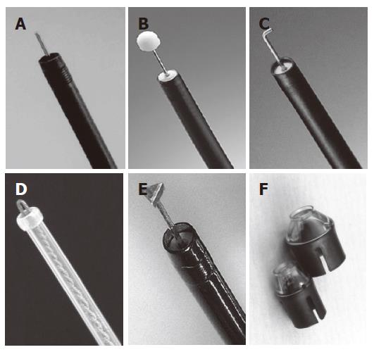
Figure 1 Devices for ESD.
A: Needle knife (KD-1L-1, Olympus, Tokyo, Japan); B: IT (KD-610L, Olympus, Tokyo, Japan); C: Hook knife (KD-620LR, Olympus, Tokyo, Japan); D: Flex knife (KD-630L, Olympus, Tokyo, Japan); E: TT knife (KD-640L, Olympus, Tokyo, Japan); F: ST hood (DH-15GR, 15CR, Fujinon Toshiba ES Systems, Tokyo, Japan).
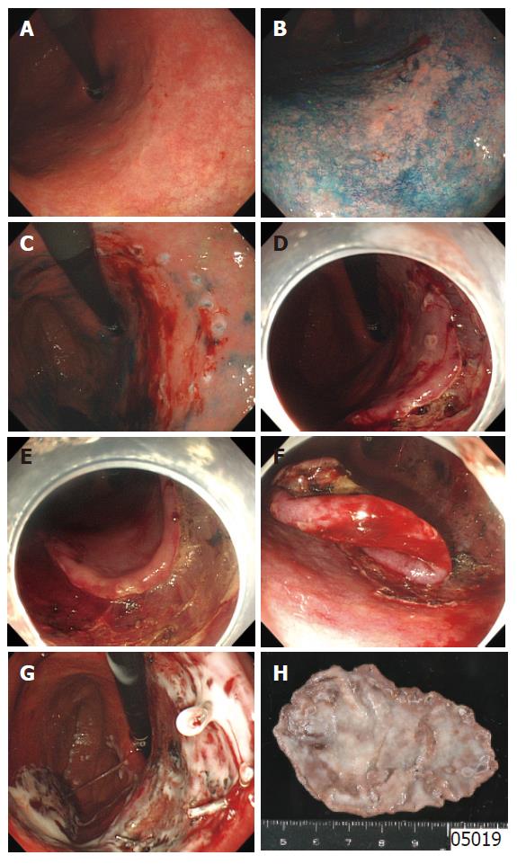
Figure 2 Endoscopic submucosal dissection (ESD).
A: Ordinal endoscopy showing a whitish slight elevation with a blurred margin in the lesser curvature of the middle gastric body; B: Chromoendoscopy revealing margins of the lesion clearly; C: Marking dots on the circumference of the lesion; D: The incised mucosa around the marking dots of the distal margins; E: Before completion of circumferential mucosal incision, submucosal dissection from the distal edges; F: After mucosal incision with slight submucosal dissection circumferentially, submucosal dissection from the edge of the posterior wall to the anterior wall; G: Complete detachment of the lesion from the muscle layer and spraying sucralfate for confirmation of hemostasis; H: The resected specimen including the whole marking dots showing en bloc resection of the lesion.
- Citation: Fujishiro M. Endoscopic submucosal dissection for stomach neoplasms. World J Gastroenterol 2006; 12(32): 5108-5112
- URL: https://www.wjgnet.com/1007-9327/full/v12/i32/5108.htm
- DOI: https://dx.doi.org/10.3748/wjg.v12.i32.5108


