Copyright
©2006 Baishideng Publishing Group Co.
World J Gastroenterol. Jan 21, 2006; 12(3): 493-495
Published online Jan 21, 2006. doi: 10.3748/wjg.v12.i3.493
Published online Jan 21, 2006. doi: 10.3748/wjg.v12.i3.493
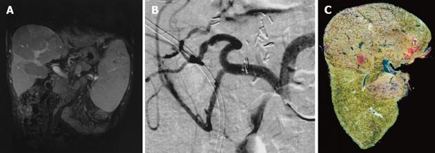
Figure 1 Pre-TIPSS MRI/angiography and post-TIPSS macroscopy of the liver.
Pictures illustrating pre-TIPSS inhomogeneous intrahepatic parenchyma texture (A); clinically irrelevant stenosis of the hepatic artery (B) and macroscopic aspect of the liver after TIPSS and organ failure due to substantial necrosis in areas where circulation was compromised before TIPSS (C).
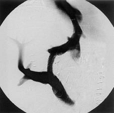
Figure 2 TIPSS-placement.
Picture illustrating correctly placed TIPSS.
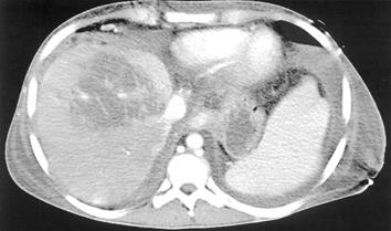
Figure 3 Spiral-CT scan after TIPSS was established.
Spiral-CT after TIPSS revealed substantial necrosis at the time of liver failure.
- Citation: Schemmer P, Radeleff B, Flechtenmacher C, Mehrabi A, Richter GM, Büchler MW, Schmidt J. TIPSS for variceal hemorrhage after living related liver transplantation: A dangerous indication. World J Gastroenterol 2006; 12(3): 493-495
- URL: https://www.wjgnet.com/1007-9327/full/v12/i3/493.htm
- DOI: https://dx.doi.org/10.3748/wjg.v12.i3.493


