Copyright
©2006 Baishideng Publishing Group Co.
World J Gastroenterol. Jan 21, 2006; 12(3): 453-456
Published online Jan 21, 2006. doi: 10.3748/wjg.v12.i3.453
Published online Jan 21, 2006. doi: 10.3748/wjg.v12.i3.453
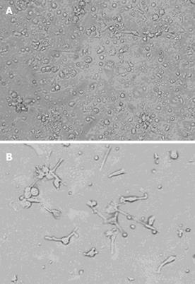
Figure 1 Dendritic cells in liquid cultures of complete RPMI 1640 supplemented with FCS, rhGM-CSF and rhIL-4.
A: Low-power view of adherent PBMC after 3 d of culture (original magnification×200). Red arrows indicate the cellular aggregates and black arrows indicate veil- or sheet-like processes of dendritic cells; B: Veil- or sheet-like processes of most cells after 7 d of culture (original magnification×400).
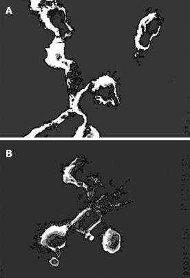
Figure 2 Cells cultivated with 10% FCS-RPMI 1640 supplemented with rhGM-CSF and rhIL-4 for 7 d (original magnification×1500).
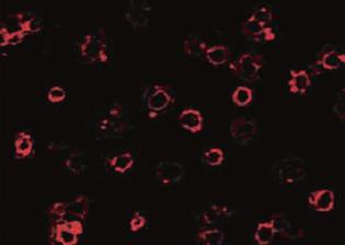
Figure 3 Cells stained with monoclonal anti-human DC-SIGN phycoerythrin (original magnification×400).
- Citation: Li J, Feng ZH, Li GY, Mou DL, Nie QH. Expression of dendritic cell-specific intercellular adhesion molecule 3 grabbing nonintegrin on dendritic cells generated from human peripheral blood monocytes. World J Gastroenterol 2006; 12(3): 453-456
- URL: https://www.wjgnet.com/1007-9327/full/v12/i3/453.htm
- DOI: https://dx.doi.org/10.3748/wjg.v12.i3.453


