Copyright
©2006 Baishideng Publishing Group Co.
World J Gastroenterol. Jul 28, 2006; 12(28): 4511-4516
Published online Jul 28, 2006. doi: 10.3748/wjg.v12.i28.4511
Published online Jul 28, 2006. doi: 10.3748/wjg.v12.i28.4511
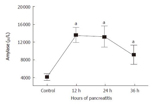
Figure 1 Serum amylase activity in pancreatitis rats (12, 24, 36 h) vs sham operated and normal controls.
aP < 0.05.
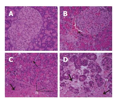
Figure 2 HE stain of normal rat pancreas (A) and after tauro-cholate injection at 12, 24, and 36 h (B, C, and D).
Induction of pancreatitis resulted in hemorrhage (thin arrows), infiltration of neutrophils (“n”), and necrosis (thick arrows).
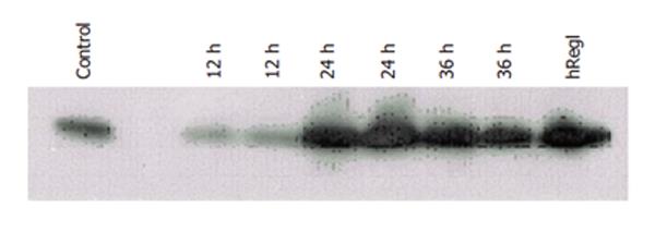
Figure 3 Slot blot analysis with anti-reg I antibody of total protein (10 μg/well) from normal rat pancreas and rat pancreas after induction of acute pancreatitis.
RegIprotein levels in tissue increased after 24 h of pancreatitis. Human RegI(hReg1) is shown as a loading control as described in MATERIALS AND METHODS.
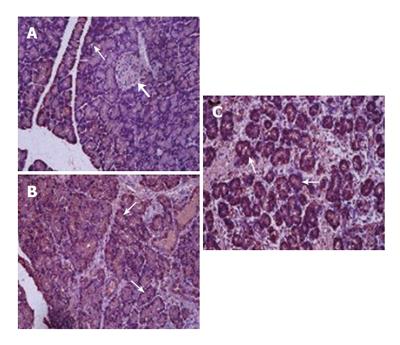
Figure 4 Immunohistochemical staining of normal rat pancreas with anti-reg antibody (A) and of rat pancreas after 24 and 36 h of pancreatitis (B, C).
There is a marked increase in regIlevels in acinar cells of the pancreas after pancreatitis. Stained acinar cells are marked with thin arrows, compared with lack of staining in Islet cells (thick arrow).
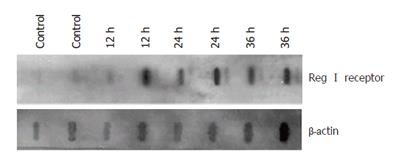
Figure 5 Slot blot of regIreceptor during pancreatitis showed an increase in regIreceptor mRNA expression after induction of pancreatitis.
The density of each reg band was normalized to that of β-actin band for the same sample as described in MATERIALS AND METHODS.
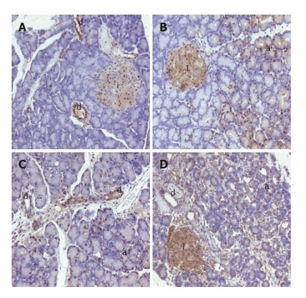
Figure 6 Immunohistochemical staining of pancreata from normal (A) and experimental pancreatitis (B-D) rats.
There is an induction of regIreceptor expression in the acinar cells (a) and islets (i), and maintenance of regIreceptor expression in ductal cells (d) with pancreatitis.
- Citation: Bluth MH, Patel SA, Dieckgraefe BK, Okamoto H, Zenilman ME. Pancreatic regenerating protein (reg I) and reg I receptor mRNA are upregulated in rat pancreas after induction of acute pancreatitis. World J Gastroenterol 2006; 12(28): 4511-4516
- URL: https://www.wjgnet.com/1007-9327/full/v12/i28/4511.htm
- DOI: https://dx.doi.org/10.3748/wjg.v12.i28.4511


