
















©2006 Baishideng Publishing Group Co.
World J Gastroenterol. Jul 28, 2006; 12(28): 4511-4516
Published online Jul 28, 2006. doi: 10.3748/wjg.v12.i28.4511
Published online Jul 28, 2006. doi: 10.3748/wjg.v12.i28.4511
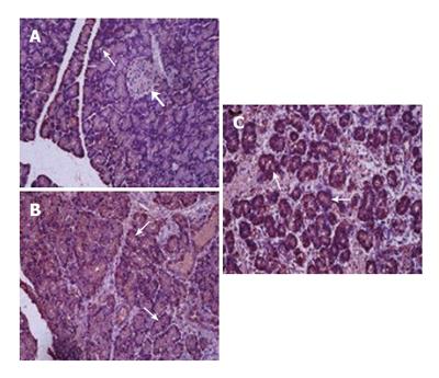
Figure 4 Immunohistochemical staining of normal rat pancreas with anti-reg antibody (A) and of rat pancreas after 24 and 36 h of pancreatitis (B, C).
There is a marked increase in regIlevels in acinar cells of the pancreas after pancreatitis. Stained acinar cells are marked with thin arrows, compared with lack of staining in Islet cells (thick arrow).
- Citation: Bluth MH, Patel SA, Dieckgraefe BK, Okamoto H, Zenilman ME. Pancreatic regenerating protein (reg I) and reg I receptor mRNA are upregulated in rat pancreas after induction of acute pancreatitis. World J Gastroenterol 2006; 12(28): 4511-4516
- URL: https://www.wjgnet.com/1007-9327/full/v12/i28/4511.htm
- DOI: https://dx.doi.org/10.3748/wjg.v12.i28.4511