Copyright
©2006 Baishideng Publishing Group Co.
World J Gastroenterol. Jul 7, 2006; 12(25): 4089-4092
Published online Jul 7, 2006. doi: 10.3748/wjg.v12.i25.4089
Published online Jul 7, 2006. doi: 10.3748/wjg.v12.i25.4089
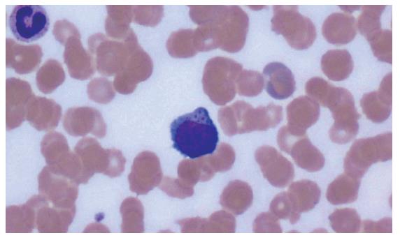
Figure 1 Peripheral blood smear-high power (1000 ×) view of atypically large lymphoid cells with blastic chromatin and abundant cytoplasm containing fine azurophilic granules.
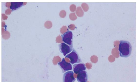
Figure 2 Ascites cell-block- high power (600 ×) view of large lymphocytes with molding, convoluted nuclear membranes, dense chromatin, and abundant cytoplasm.
Features are supportive of possible T-cell morphology.
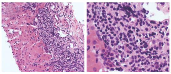
Figure 3 Liver biopsy-low power (200 ×) view of the liver parenchyma infiltrated by a malignant lymphoid population composed of intermediate-sized to large atypical lymphocytes (A) and liver biopsy-high power (1000 ×) view of the liver parenchyma showing intermediate-sized to large atypical lymphocytes with very irregular nuclear contours and dense nuclear chromatin (B).
- Citation: Dellon ES, Morris SR, Tang W, Dunphy CH, Russo MW. Acute liver failure due to natural killer-like T-cell leukemia/lymphoma: A case report and review of the Literature. World J Gastroenterol 2006; 12(25): 4089-4092
- URL: https://www.wjgnet.com/1007-9327/full/v12/i25/4089.htm
- DOI: https://dx.doi.org/10.3748/wjg.v12.i25.4089


