Copyright
©2006 Baishideng Publishing Group Co.
World J Gastroenterol. Jun 28, 2006; 12(24): 3933-3935
Published online Jun 28, 2006. doi: 10.3748/wjg.v12.i24.3933
Published online Jun 28, 2006. doi: 10.3748/wjg.v12.i24.3933
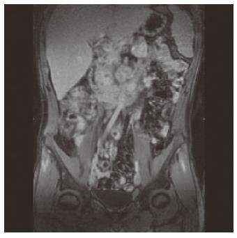
Figure 1 MRI coronal view, T1-weighted post oral administration of super-paramagnetic contrast agent.
Image illustrates marked wall thickening and enhancement at the level of terminal ileum and enlarged mesenteric lymph nodes.
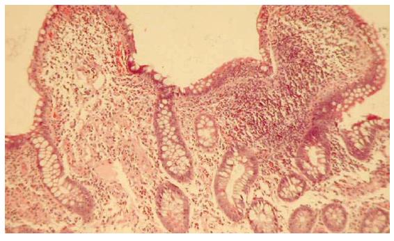
Figure 2 Biopsy of the terminal ileum showing histopathological findings of Crohn’s disease.
- Citation: Zippi M, Colaiacomo MC, Marcheggiano A, Pica R, Paoluzi P, Iaiani G, Caprilli R, Maccioni F. Mesenteric adenitis caused by Yersinia pseudotubercolosis in a patient subsequently diagnosed with Crohn’s disease of the terminal ileum. World J Gastroenterol 2006; 12(24): 3933-3935
- URL: https://www.wjgnet.com/1007-9327/full/v12/i24/3933.htm
- DOI: https://dx.doi.org/10.3748/wjg.v12.i24.3933


