Copyright
©2006 Baishideng Publishing Group Co.
World J Gastroenterol. Jun 28, 2006; 12(24): 3929-3932
Published online Jun 28, 2006. doi: 10.3748/wjg.v12.i24.3929
Published online Jun 28, 2006. doi: 10.3748/wjg.v12.i24.3929
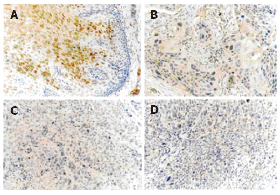
Figure 1 Immunohistochemical analysis of TGM3 in paired ESCC tissue samples using anti- TGM3 antibody (1:100).
Diffuse and strong staining was detected in the cytoplasm of the normal epithelial cells (A), while sporadic and weak staining was observed in the cytoplasm of matched esophageal cancer epithelial cells (B: well-differentiated, C: moderately-differentiated, D: poorly-differentiated) (original magnification × 200).
- Citation: Liu W, Yu ZC, Cao WF, Ding F, Liu ZH. Functional studies of a novel oncogene TGM3 in human esophageal squamous cell carcinoma. World J Gastroenterol 2006; 12(24): 3929-3932
- URL: https://www.wjgnet.com/1007-9327/full/v12/i24/3929.htm
- DOI: https://dx.doi.org/10.3748/wjg.v12.i24.3929


