Copyright
©2006 Baishideng Publishing Group Co.
World J Gastroenterol. Jan 14, 2006; 12(2): 317-321
Published online Jan 14, 2006. doi: 10.3748/wjg.v12.i2.317
Published online Jan 14, 2006. doi: 10.3748/wjg.v12.i2.317
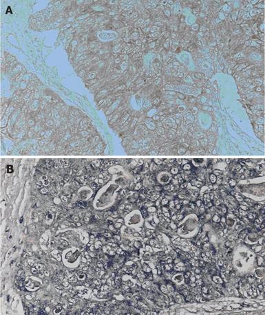
Figure 1 Immunohistochemical staining for the IL-11 and IL-11Rα in human colorectal carcinoma.
(A) is for IL-11, and (B) is for IL-11Rα. IL-11 and IL-11Rα show strong cytoplasmic and membranous expression.
Figure 2 Demonstration of IL-11 and IL-11Rα in colorectal carcinoma cells and tissues by Western blotting.
All of colorectal carcinoma cells, carcinoma tissues and normal mucosa expressed both IL-11 and IL-11Rα. (N: normal mucosa, T: human colorectal cancer tissue).
- Citation: Yamazumi K, Nakayama T, Kusaba T, Wen CY, Yoshizaki A, Yakata Y, Nagayasu T, Sekine I. Expression of Interleukin-11 and Interleukin-11 receptor α in human colorectal adenocarcinoma; Immunohistochemical analyses and correlation with clinicopathological factors. World J Gastroenterol 2006; 12(2): 317-321
- URL: https://www.wjgnet.com/1007-9327/full/v12/i2/317.htm
- DOI: https://dx.doi.org/10.3748/wjg.v12.i2.317


