Copyright
©2006 Baishideng Publishing Group Co.
World J Gastroenterol. May 7, 2006; 12(17): 2762-2766
Published online May 7, 2006. doi: 10.3748/wjg.v12.i17.2762
Published online May 7, 2006. doi: 10.3748/wjg.v12.i17.2762
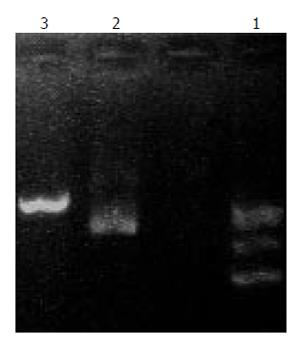
Figure 2 pcDNA3.
1(+)-angiostatin linearization. Lane 1: λDNA/HindIII marker; lane 2: pcDNA3.1(+)-angiostatin; lane 3: pcDNA3.1(+)-angiostatin /PvuI.
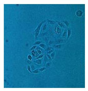
Figure 3 Gallbladder carcinoma cell clones transfected with pcDNA3.
1(+)-angiostatin.× 400.
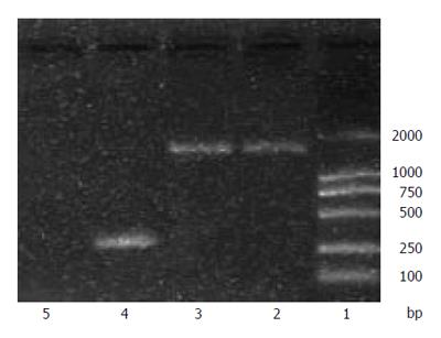
Figure 4 Analysis of angiostatin transcript by RT-PCR.
Lane1: DNA marker DL2000; lanes 2, 3: angiostatin; lane 4: positive control; lane 5: nagetive control.
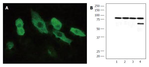
Figure 5 Angiostatin protein expression by immunofluorescence(A) and Western blot analysis(B).
M:Marker; lane 1: PBS control; lanes 2,3: GBC-SDpcDNA3.1(+); lane 4: GBC-SDpcDNA(3.1)angiostatin.
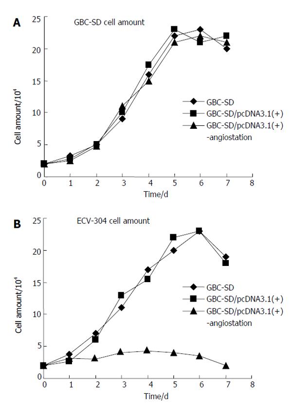
Figure 6 Cell growth curves of GBC-SD(A) and ECV-304(B).
- Citation: Yang DZ, He J, Zhang JC, Wang ZR. Expression of angiostatin cDNA in human gallbladder carcinoma cell line GBC-SD and its effect on endothelial proliferation and growth. World J Gastroenterol 2006; 12(17): 2762-2766
- URL: https://www.wjgnet.com/1007-9327/full/v12/i17/2762.htm
- DOI: https://dx.doi.org/10.3748/wjg.v12.i17.2762


