Copyright
©2006 Baishideng Publishing Group Co.
World J Gastroenterol. Apr 28, 2006; 12(16): 2477-2486
Published online Apr 28, 2006. doi: 10.3748/wjg.v12.i16.2477
Published online Apr 28, 2006. doi: 10.3748/wjg.v12.i16.2477
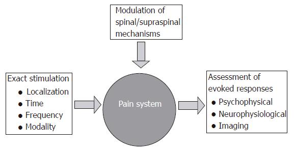
Figure 1 The concept for experimental induction, assessment and modulation of experimental gut pain in man.
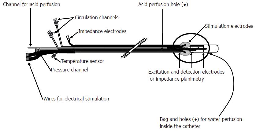
Figure 2 Schematic illustration of the probe used for multi-modal (electrical, mechanical, cold and warmth stimuli) of the esophagus.
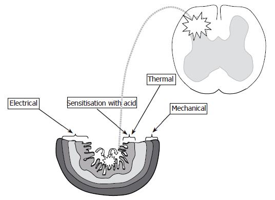
Figure 3 Schematic illustration of the proposed gut layers which are preferentially affected with: (1) Thermal stimuli (mucosa - dark grey and submucosa - light grey); (2) Mechanical stimuli (circular muscle layer - hatched grey and longitudinal muscle layer - prickled grey); (3) Electrical stimuli (all layers depending on stimulus intensity).
Perfusion of the esophagus with acid (curved arrows) gives the possibility to evoke peripheral and (mainly) central sensitization (illustrated with stars).
Figure 4 Cross-sectional area (CSA) at moderate pain during two distensions of the esophagus in a patient with non-cardiac chest pain.
The distensions were performed before and after perfusion of the distal esophagus with acid (illustrated with the stippled line). A clear reduction in the tolerated mechanical stimulus was seen after acid perfusion.
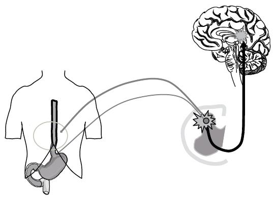
Figure 5 Referred pain in the somatic tissues is believed to be generated by central mechanisms, where visceral and somatic nerves converge on nerves in the same area of the spinal cord or at supraspinal centers.
The phenomena also includes unmasking of latent connections and focal central hyperexcitability of the neurons.
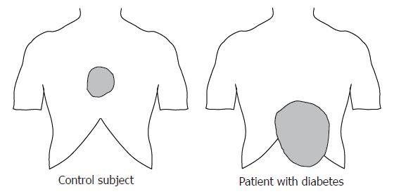
Figure 6 Referred pain area to mechanical distension of the esophagus in a typical healthy subject, and in a patient with diabetes mellitus and autonomic neuropathy.
The patient complained of severe nausea and pain in the epigastrium. The referred pain area in the diabetic patient was larger and abnormally localized.
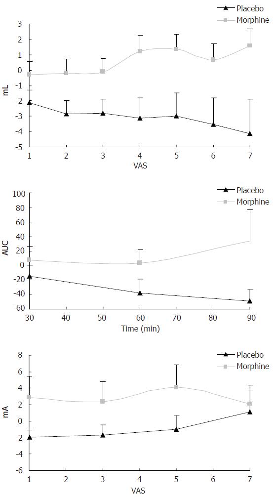
Figure 7 Change in sensory rating compared with baseline recordings for oral morphine and placebo using multimodal stimulation of the esophagus.
Top picture: Morphine attenuates mechanically evoked pain better than placebo 60 min after drug intake (P < 0.01). VAS on the X-axis denotes the sensory rating at a visual analogue scale with 5 as the pain threshold. ML at the Y-axis denotes the bag volume. Middle picture: Morphine attenuates heat pain better than placebo (P < 0.05). Here the X-axis illustrates time after drug intake (min) and AUC on the Y-axis denotes the area under the temperature curve used as a proxy for the thermal energy. Bottom picture: Morphine also worked better than placebo (P < 0.05) on electrical stimulation 60 min after drug intake.
- Citation: Drewes AM, Gregersen H. Multimodal pain stimulation of the gastrointestinal tract. World J Gastroenterol 2006; 12(16): 2477-2486
- URL: https://www.wjgnet.com/1007-9327/full/v12/i16/2477.htm
- DOI: https://dx.doi.org/10.3748/wjg.v12.i16.2477


