Copyright
©2006 Baishideng Publishing Group Co.
World J Gastroenterol. Apr 21, 2006; 12(15): 2445-2449
Published online Apr 21, 2006. doi: 10.3748/wjg.v12.i15.2445
Published online Apr 21, 2006. doi: 10.3748/wjg.v12.i15.2445
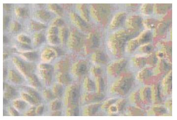
Figure 1 A phase-contrast micrograph of normal human esopha-geal epithelial cells (×250).
Figure 2 Effect of acid exposure on cell proliferation.
Figure 3 Effect of bile acid exposure on cell proliferation.
Figure 4 Changes in proliferation of cells exposed to bile acid with different pH for 3 min.
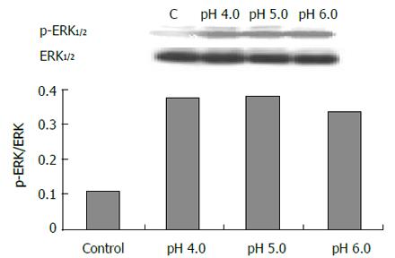
Figure 5 Effect of acid exposure (3 min) on ERK.
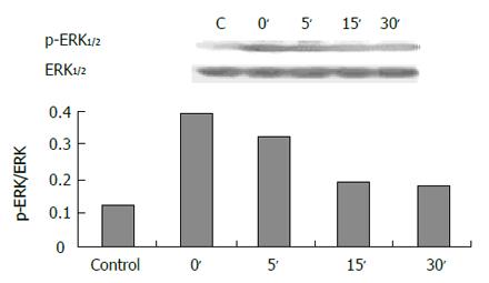
Figure 6 Expression of ERK in 30 min after acid exposure (pH 4.
0, 3 min).
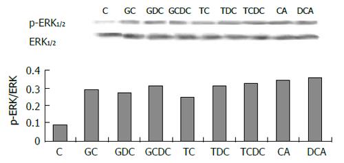
Figure 7 Effect of bile acid exposure (3 min) on ERK.
- Citation: Jiang ZR, Gong J, Zhang ZN, Qiao Z. Influence of acid and bile acid on ERK activity, PPARγ expression and cell proliferation in normal human esophageal epithelial cells. World J Gastroenterol 2006; 12(15): 2445-2449
- URL: https://www.wjgnet.com/1007-9327/full/v12/i15/2445.htm
- DOI: https://dx.doi.org/10.3748/wjg.v12.i15.2445


