Copyright
©2006 Baishideng Publishing Group Co.
World J Gastroenterol. Apr 21, 2006; 12(15): 2341-2344
Published online Apr 21, 2006. doi: 10.3748/wjg.v12.i15.2341
Published online Apr 21, 2006. doi: 10.3748/wjg.v12.i15.2341
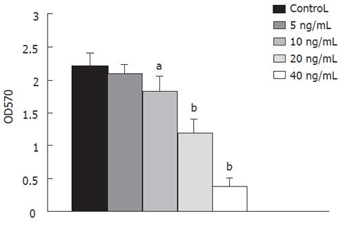
Figure 1 Effect of different concentrations of StxA1-GM-CSF on LS174T cell line by MTT assay.
(aP < 0.01, bP < 0.001 vs trypan blue exclusion assay).
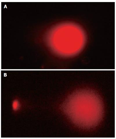
Figure 2 Morphology of normal cells (A) and apoptotic cells (B) after treatment with hybrid protein.
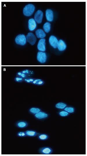
Figure 3 Hoechst 33342-staining of LS174T cells treated with StxA1-GM-CSF.
A: control; B: cells after treatment.
Figure 4 Apoptosis percentage in the presence of 40 ng/mL StxA1-GM-CSF at different time intervals (A), aP < 0.
05, bP < 0.001. Apoptosis percentage in the presence of different concentration of StxA1-GMCSF for 6 h (B). dP < 0.01, fP < 0.001.
- Citation: Roudkenar MH, Bouzari S, Kuwahara Y, Roushandeh AM, Oloomi M, Fukumoto M. Recombinant hybrid protein, Shiga toxin and granulocyte macrophage colony stimulating factor effectively induce apoptosis of colon cancer cells. World J Gastroenterol 2006; 12(15): 2341-2344
- URL: https://www.wjgnet.com/1007-9327/full/v12/i15/2341.htm
- DOI: https://dx.doi.org/10.3748/wjg.v12.i15.2341


