Copyright
©2006 Baishideng Publishing Group Co.
World J Gastroenterol. Apr 21, 2006; 12(15): 2335-2340
Published online Apr 21, 2006. doi: 10.3748/wjg.v12.i15.2335
Published online Apr 21, 2006. doi: 10.3748/wjg.v12.i15.2335
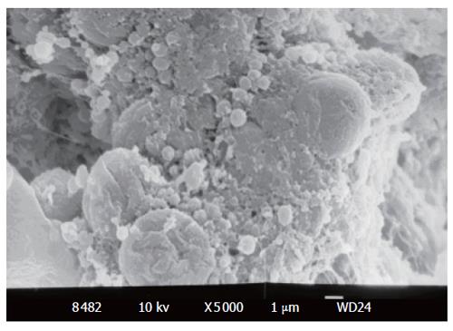
Figure 1 Cocci in adjacent hepatocytes (SEM × 5000).
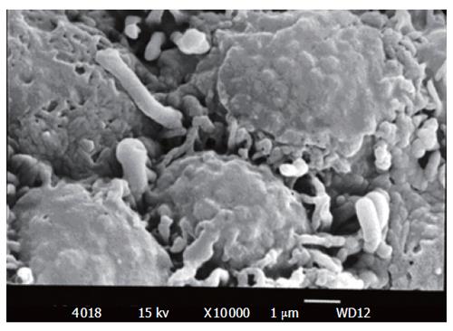
Figure 2 Spiral-shaped bacteria in vicinity of pit cell lineage (SEM × 10 000).
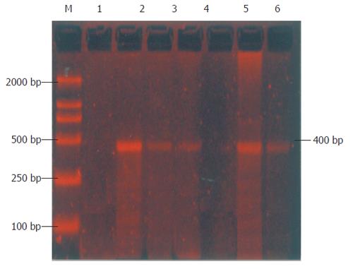
Figure 3 Analysis of Helicobacter spp.
PCR products from HCC samples. The 400-bp fragments were analyzed by 1.5% agarose gel electrophoresis. Lane M: nucleotide marker; lane 1: negative control (double-distilled water); lane 2: positive control (H pylori DNA); Lanes 3, 4, 6: positive samples.
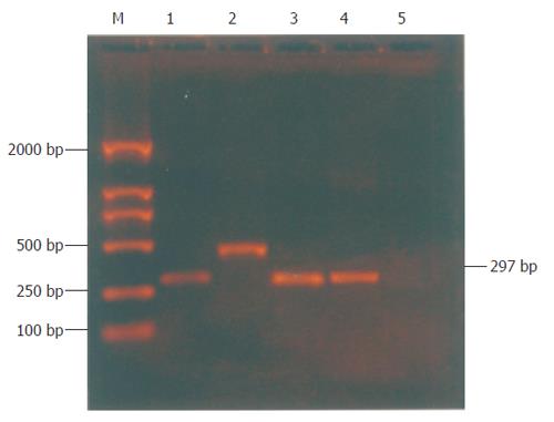
Figure 4 Analysis of vacA and cagA PCR products from HCC samples.
The 352-bp and 297-bp fragments were analyzed by 1.5% agarose gel electrophoresis. Lane M: nucleotide marker; lane 1: positive control (cagA DNA); lane 2: positive control (vacA DNA); Lanes 3, 4: cagA positive samples; lane 5: negative control (double-distilled water).
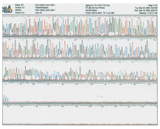
Figure 5 Sequencing results of 16S rRNA in helicobacter genus-positive products from HCC.
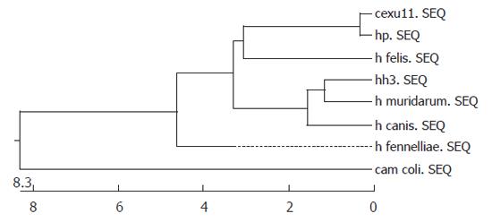
Figure 6 Genic phylogenetic tree of Helicobacter spp.
and deduced protein.
- Citation: Xuan SY, Li N, Qiang X, Zhou RR, Shi YX, Jiang WJ. Helicobacter infection in hepatocellular carcinoma tissue. World J Gastroenterol 2006; 12(15): 2335-2340
- URL: https://www.wjgnet.com/1007-9327/full/v12/i15/2335.htm
- DOI: https://dx.doi.org/10.3748/wjg.v12.i15.2335


