Copyright
©2006 Baishideng Publishing Group Co.
World J Gastroenterol. Apr 14, 2006; 12(14): 2305-2307
Published online Apr 14, 2006. doi: 10.3748/wjg.v12.i14.2305
Published online Apr 14, 2006. doi: 10.3748/wjg.v12.i14.2305
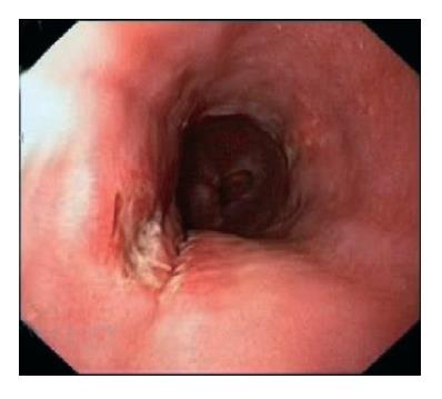
Figure 1 Exudate-covered ulcer in distal esophagus.
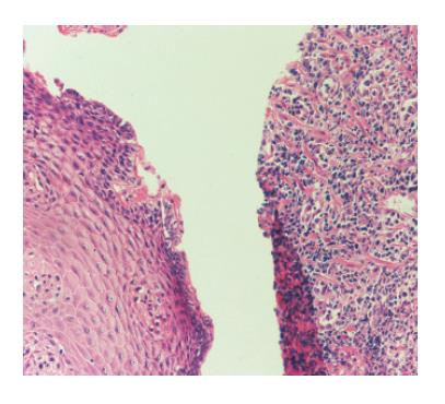
Figure 2 Plasma cells infiltrating esophageal mucosa (hematoxylin & eosin staining, x 200).
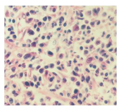
Figure 3 Myeloma cells in lamina propria of esophagus (hematoxylin & eosin staining, x 400).
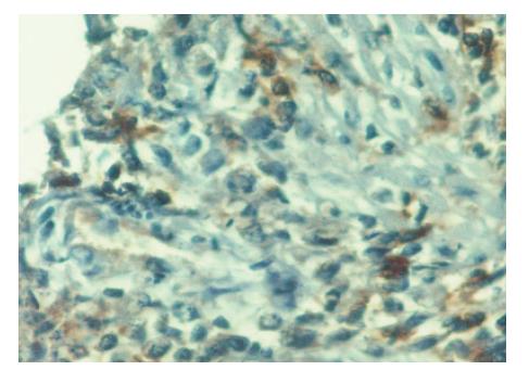
Figure 4 Immunohistochemically stained kappa light-chain showing monoclonality of plasma cells (x400).
- Citation: Pehlivan Y, Sevinc A, Sari I, Gulsen MT, Buyukberber M, Kalender ME, Camci C. An interesting cause of esophageal ulcer etiology: Multiple myeloma of IgG kappa subtype. World J Gastroenterol 2006; 12(14): 2305-2307
- URL: https://www.wjgnet.com/1007-9327/full/v12/i14/2305.htm
- DOI: https://dx.doi.org/10.3748/wjg.v12.i14.2305


