Copyright
©2006 Baishideng Publishing Group Co.
World J Gastroenterol. Apr 7, 2006; 12(13): 2130-2132
Published online Apr 7, 2006. doi: 10.3748/wjg.v12.i13.2130
Published online Apr 7, 2006. doi: 10.3748/wjg.v12.i13.2130
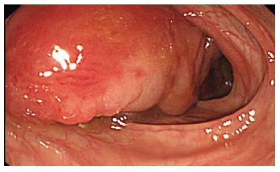
Figure 1 Colonoscopic examination shows a 2.
5 cm x 2.5 cm x 5.0 cm sized round, smooth surfaced mass covered by normal mucosa in the descending colon. This mass is pedunclated cystic nature, and compressible pillow sign positive.
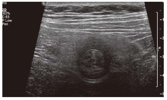
Figure 2 Ultrasonography of the mass shows a multilocular cystic lesion with septa.
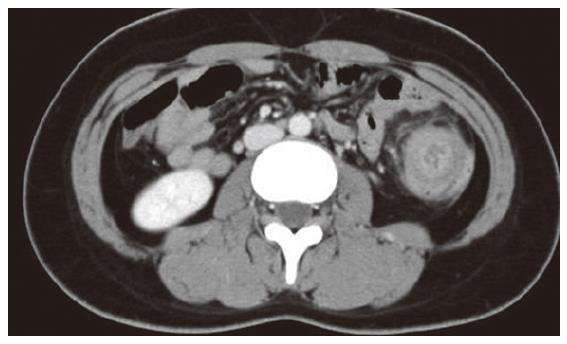
Figure 3 Computed tomography shows the cystic lesion in the descending colon, occupying the whole lumen, and also shows multicentric target sign and sausage-shaped inhomogeneous soft-tissue mass.
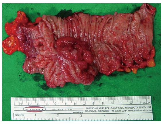
Figure 4 Macroscopic finding of the resected specimen reveals 2.
5 cm x 3.5 cm x 5.0 cm sized multiseptated submucosal cystic tumor, which changed shape easily. The tumor shows fluctuation and serous clear fluid was aspirated by needle puncture.
Figure 5 (A) Cystically dilated spaces are covered with single layer of endothelial cells and separated by fibrous septa are present in the submucosa.
The overlying colonic mucosa is normal (H & E stain, x100). (B) Atypical cell are not noted, and endothelium is lined by benign cuboidal cells (H-E, original magnification, x400).
- Citation: Kim TO, Lee JH, Kim GH, Heo J, Kang DH, Song GA, Cho M. Adult intussusception caused by cystic lymphangioma of the colon: A rare case report. World J Gastroenterol 2006; 12(13): 2130-2132
- URL: https://www.wjgnet.com/1007-9327/full/v12/i13/2130.htm
- DOI: https://dx.doi.org/10.3748/wjg.v12.i13.2130


