Copyright
©2006 Baishideng Publishing Group Co.
World J Gastroenterol. Mar 28, 2006; 12(12): 1937-1940
Published online Mar 28, 2006. doi: 10.3748/wjg.v12.i12.1937
Published online Mar 28, 2006. doi: 10.3748/wjg.v12.i12.1937
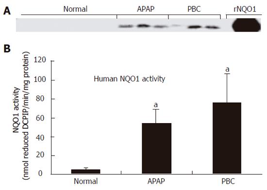
Figure 1 NQO1 protein (A) and activity (B) in cytosolic fractions from normal, APAP and PBC human liver specimens.
The data are presented as nmol reduced DCPIP/min/mg protein ± SE (n = 3-5, aP < 0.05 vs normal liver specimens).
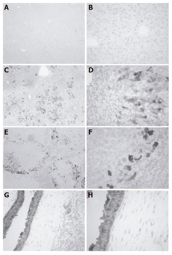
Figure 2 Immunoperoxidase staining of NQO1 in normal (A, B), APAP (C, D), and PBC (E, F) human liver specimens as well as in PBC hyperplastic biliary epithelium (G, H).
- Citation: Aleksunes LM, Goedken M, Manautou JE. Up-regulation of NAD(P)H quinone oxidoreductase 1 during human liver injury. World J Gastroenterol 2006; 12(12): 1937-1940
- URL: https://www.wjgnet.com/1007-9327/full/v12/i12/1937.htm
- DOI: https://dx.doi.org/10.3748/wjg.v12.i12.1937


