Copyright
©2005 Baishideng Publishing Group Inc.
World J Gastroenterol. Mar 7, 2005; 11(9): 1399-1402
Published online Mar 7, 2005. doi: 10.3748/wjg.v11.i9.1399
Published online Mar 7, 2005. doi: 10.3748/wjg.v11.i9.1399
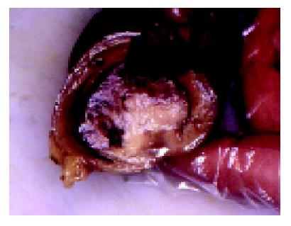
Figure 1 Polypoid tumor in intestinal lumen.
On the cut surface the tumor was whitish, flashy and soft.
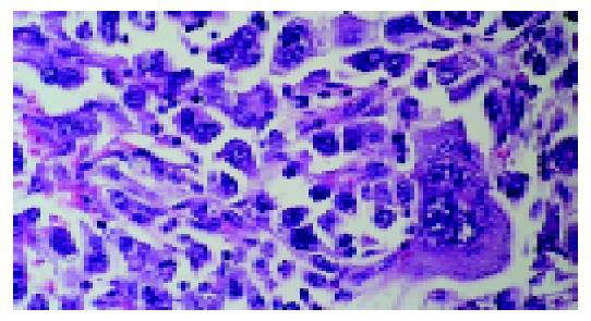
Figure 2 Tumor consisted almost exclusively of strands and sheets of poorly cohesive, polymorphic giant cells with scanty, delicate stromas (hematoxylin-eosin ×400).
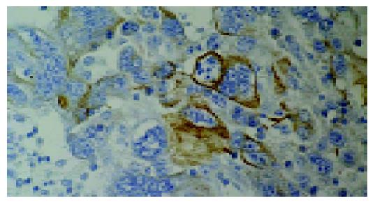
Figure 3 Focal positivity for pancytokeratin in tumor cells(MSIP ×400).
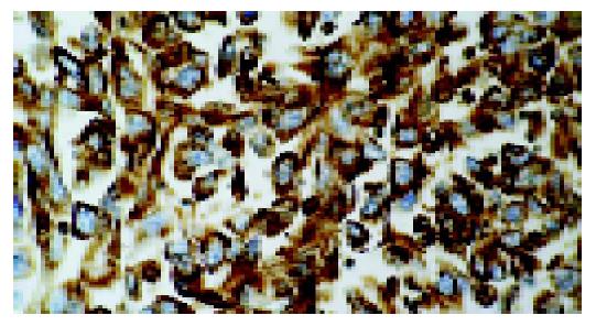
Figure 4 Diffuse intense staining for vimentin in tumor cells (MSIP ×400).
- Citation: Tomas D, Ledinsky M, Belicza M, Krušlin B. Multiple metastases to the small bowel from large cell bronchial carcinomas. World J Gastroenterol 2005; 11(9): 1399-1402
- URL: https://www.wjgnet.com/1007-9327/full/v11/i9/1399.htm
- DOI: https://dx.doi.org/10.3748/wjg.v11.i9.1399


