Copyright
©2005 Baishideng Publishing Group Inc.
World J Gastroenterol. Feb 7, 2005; 11(5): 764-766
Published online Feb 7, 2005. doi: 10.3748/wjg.v11.i5.764
Published online Feb 7, 2005. doi: 10.3748/wjg.v11.i5.764
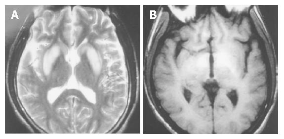
Figure 1 Cranial MRI in the patient with similar hepatolenticular degeneration (Wilson’s disease).
A: increased signal intensity in basal ganglia bilaterally on T2WI; B: increased signal intensity in basal ganglia bilaterally on T1WI.
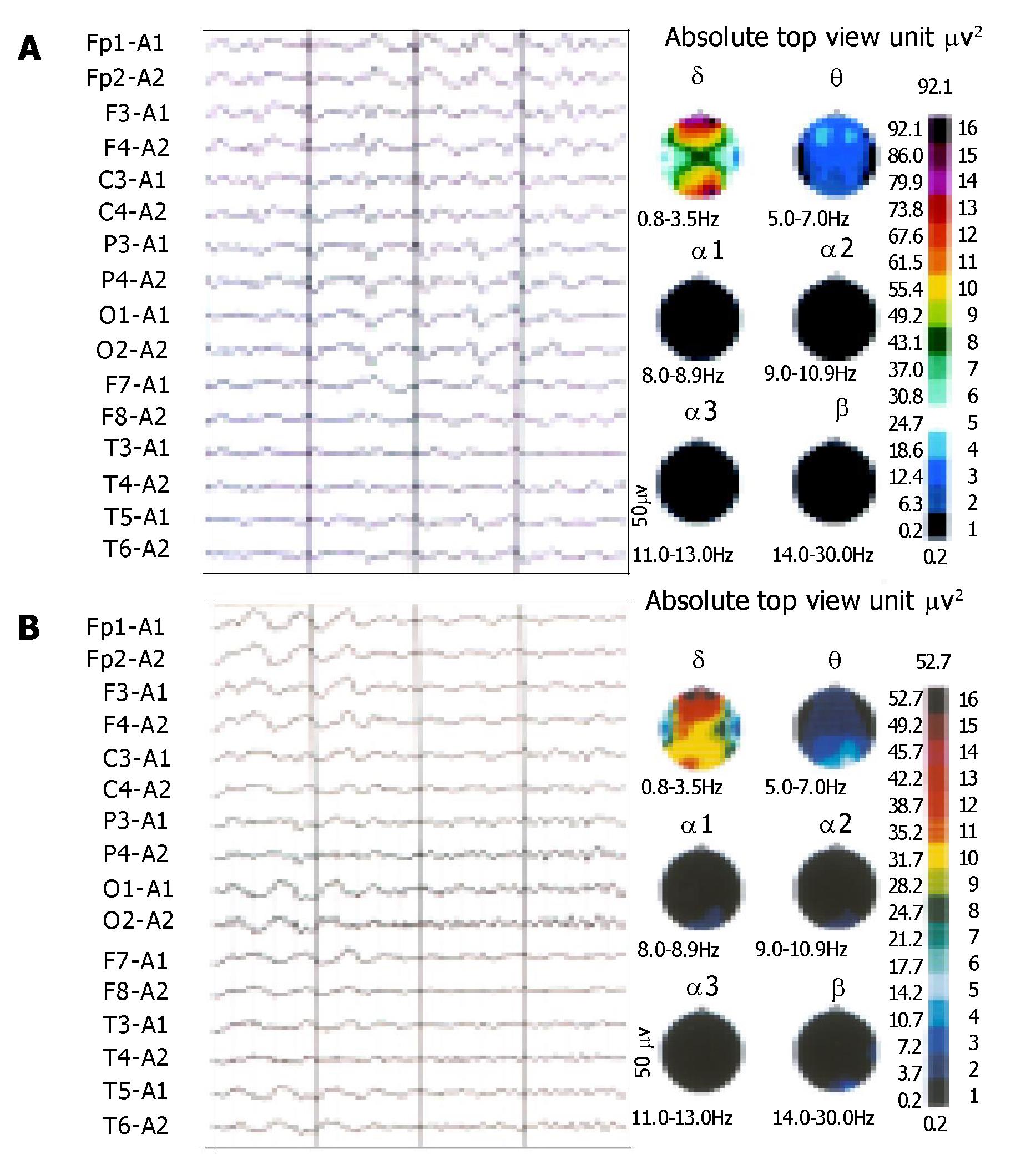
Figure 2 θ wave intermixed with δ wave, triphasic wave and spike and ware wave on EEG before treatmant(A).
and waves reduction on EEG after treatmant(B).
- Citation: Chen WX, Wang P, Yan SX, Li YM, Yu CH, Jiang LL. Acquired hepatocerebral degeneration: A case report. World J Gastroenterol 2005; 11(5): 764-766
- URL: https://www.wjgnet.com/1007-9327/full/v11/i5/764.htm
- DOI: https://dx.doi.org/10.3748/wjg.v11.i5.764


