Copyright
©2005 Baishideng Publishing Group Inc.
World J Gastroenterol. Nov 14, 2005; 11(42): 6644-6649
Published online Nov 14, 2005. doi: 10.3748/wjg.v11.i42.6644
Published online Nov 14, 2005. doi: 10.3748/wjg.v11.i42.6644
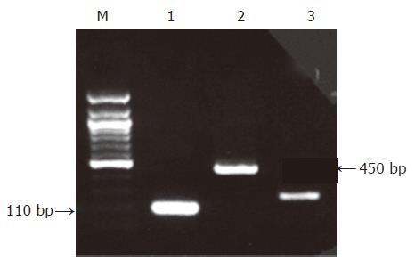
Figure 1 Nested PCR with HSV-1 outer and inner primers (450- and 110-bp amplicons: arrows) on 2% agarose gel, by ethidium bromide staining.
Lane M: DNA molecular weight marker Φ×174/Hae III, lane 1: HSV-1 positive control (second run of nested PCR), lane 2: HSV-1 positive control (first run of nested PCR), lane 3: b-actin quality control PCR peptic ulcer sample.
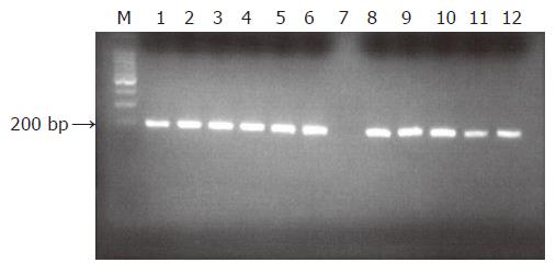
Figure 2 PCR quality assay control.
Lane M: DNA molecular weight marker Φ×174/Hae III, lanes 1-6: positive b-actin peptic ulcer samples; lane 7: negative control (without template); lanes 8-12: positive b-actin peptic ulcer samples.
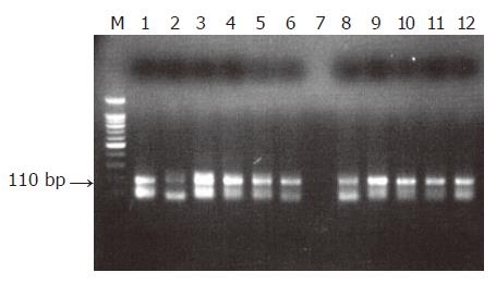
Figure 3 Nested PCR amplification of HSV-1 in samples of patients with peptic ulcer.
Lane M: DNA molecular weight marker Φ×174/Hae III, lane 1: HSV-1 positive control (HSV-1 genomic DNA by Sigma), lanes 2-6: positive samples from patients with peptic ulcer, lane 7: negative control (without template), lanes 8-11: positive samples from patients with peptic ulcer, lane 12: HSV-1 positive control.
-
Citation: Tsamakidis K, Panotopoulou E, Dimitroulopoulos D, Xinopoulos D, Christodoulou M, Papadokostopoulou A, Karagiannis I, Kouroumalis E, Paraskevas E. Herpes simplex virus type 1 in peptic ulcer disease: An inverse association with
Helicobacter pylori . World J Gastroenterol 2005; 11(42): 6644-6649 - URL: https://www.wjgnet.com/1007-9327/full/v11/i42/6644.htm
- DOI: https://dx.doi.org/10.3748/wjg.v11.i42.6644


