Copyright
©2005 Baishideng Publishing Group Inc.
World J Gastroenterol. Nov 7, 2005; 11(41): 6560-6562
Published online Nov 7, 2005. doi: 10.3748/wjg.v11.i41.6560
Published online Nov 7, 2005. doi: 10.3748/wjg.v11.i41.6560
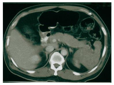
Figure 1 Abdominal computed tomography identifies an enlarged spleen with a heterogeneous density involving the whole spleen.
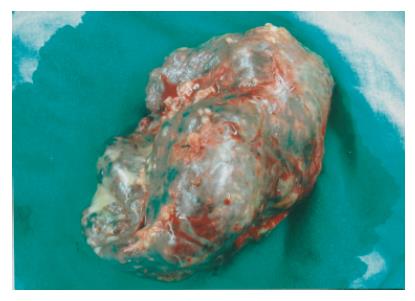
Figure 2 The splenic parenchyma is diffusely scattered with multiple, irregular sized and dark-red nodules.
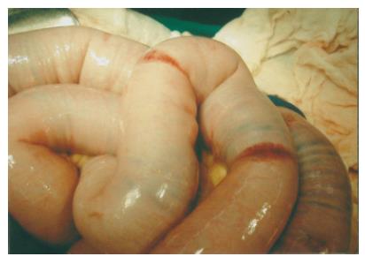
Figure 3 Multiple small hemorrhagic patches are identified at the jejunum and ileum.
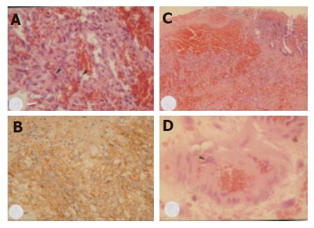
Figure 4 A: Photomicrography of the spleen reveals neoplastic sinusoidal endothelial cells with marked cytological atypia (arrowhead) lining the congested sinusoid spaces (hematoxylin and eosin staining, 200×); B: immunohistochemical staining of the neoplastic cells is positive for factor XIII associated antigen (immunostain, 200×); C: histological examination of the specimen of small intestine shows metastatic neoplastic cells primarily distributed within the submucosal layer and focally extending to the mucosal layer (hematoxylin and eosin staining, 100×); D: photomicrography of the intestinal vessel reveals presence of individual neoplastic cells (arrowhead) within the vascular lumen (hematoxylin and eosin staining, 400×).
- Citation: Hsu JT, Lin CY, Wu TJ, Chen HM, Hwang TL, Jan YY. Splenic angiosarcoma metastasis to small bowel presented with gastrointestinal bleeding. World J Gastroenterol 2005; 11(41): 6560-6562
- URL: https://www.wjgnet.com/1007-9327/full/v11/i41/6560.htm
- DOI: https://dx.doi.org/10.3748/wjg.v11.i41.6560


