Copyright
©2005 Baishideng Publishing Group Inc.
World J Gastroenterol. Nov 7, 2005; 11(41): 6554-6556
Published online Nov 7, 2005. doi: 10.3748/wjg.v11.i41.6554
Published online Nov 7, 2005. doi: 10.3748/wjg.v11.i41.6554
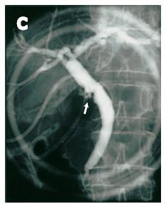
Figure 1 Endoscopic retrograde cholangiography.
The cystic duct is poorly opacified with small protruding lesions around the confluence of the cystic duct (arrow). An 8-Fr pig-tail catheter (C) is inserted into the gallbladder.
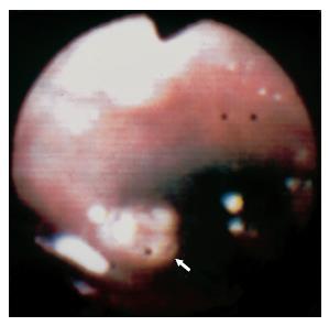
Figure 2 Peroral cholangioscopy shows a small papillary lesion (arrow) around the confluence of the cystic duct.
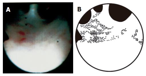
Figure 3 Peroral cholangioscopy of the interior of the right hepatic duct.
A: Fine granular mucosal lesions, brownish in color, are scattered, suggesting the presence of papillary in situ carcinoma. The flat mucosae between the fine granular lesions, which appear normal on cholangioscopy, corresponded to non-papillary in situ carcinoma histologically. The lower part of this figure is fogged owing to halation. B: Schematic representation. The dotted areas indicate the fine granular lesions.
- Citation: Wakai T, Shirai Y, Hatakeyama K. Peroral cholangioscopy for non-invasive papillary cholangiocarcinoma with extensive superficial ductal spread. World J Gastroenterol 2005; 11(41): 6554-6556
- URL: https://www.wjgnet.com/1007-9327/full/v11/i41/6554.htm
- DOI: https://dx.doi.org/10.3748/wjg.v11.i41.6554


