Copyright
©2005 Baishideng Publishing Group Inc.
World J Gastroenterol. Jan 28, 2005; 11(4): 561-566
Published online Jan 28, 2005. doi: 10.3748/wjg.v11.i4.561
Published online Jan 28, 2005. doi: 10.3748/wjg.v11.i4.561
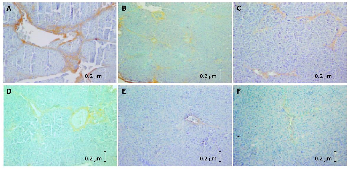
Figure 1 Expression of collagens I and III in each group (SABC×100).
A and B: In hepatic fibrosis group, a large amount of collagen fibers distributed extensively in portals, hepatic sinusoids and hepatic parenchyma, along with the formation of fibrous septa and pseudo lobules; C and D: In non-DSHX-treated group, hepatic fibrosis was lightly alleviated compared with Figures A and B, while the collagen fibers were still expanded extensively and the fibrous septa could be seen apparently; E and F: In DSHX-treated group, fewer collagen fibers were distributed in portals and expanded lightly compared with Figures C and D. No pseudo lobules formed.
Figure 2 Apoptotic index of HSCs detected by flow cytometry in each group of rats.
A: Normal group; B: Hepatic fibrosis group; C: DSHX-treated group.
-
Citation: Geng XX, Yang Q, Xie RJ, Luo XH, Han B, Ma L, Li CX, Cheng ML.
In vivo effects of Chinese herbal recipe, Danshaohuaxian, on apoptosis and proliferation of hepatic stellate cells in hepatic fibrotic rats. World J Gastroenterol 2005; 11(4): 561-566 - URL: https://www.wjgnet.com/1007-9327/full/v11/i4/561.htm
- DOI: https://dx.doi.org/10.3748/wjg.v11.i4.561


