Copyright
©2005 Baishideng Publishing Group Inc.
World J Gastroenterol. Jan 28, 2005; 11(4): 492-497
Published online Jan 28, 2005. doi: 10.3748/wjg.v11.i4.492
Published online Jan 28, 2005. doi: 10.3748/wjg.v11.i4.492
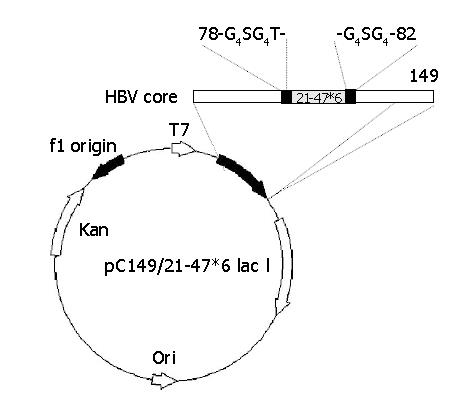
Figure 1 Construction of plasmid pC149/21-47*6.
The horizontal bar represents the chimeric HBc-preS1 (21-47) protein. The insertion foreign DNA designed such that the 21-47*6 sequence, was flanked on both sides by glycine-rich linkers, and inserted into the major immunodominant region (MIR) of truncated, assembly-competent core protein derivative core 1-149. The authentic amino acids of 79-81 were removed.
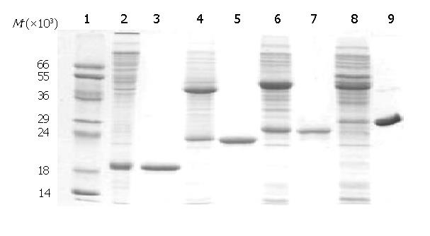
Figure 2 SDS-PAGE analysis of purified recombinant antigens.
1: protein MW marker; 2: C149/mut; 3: purified C149/mut; 4: CI; 5: purified CI; 6: CII; 7: purified CII; 8: CIII; 9: purified CIII.
Figure 3 Transmission electron microscopy of VLP antigens purified from E.
coli. The VLPs were stained with 1.5% uranyl acetate and photographed at 67000 magnification.
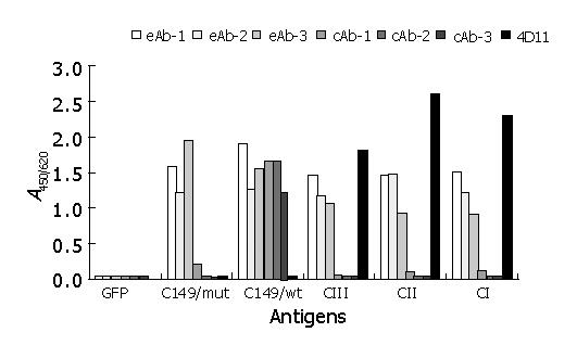
Figure 4 Reactivity of recombinant antigens with different HBV McAbs by ELISA.
GFP: cell lysates containing recombinant GFP protein at 100 mg/L as negative coating control; recombinant proteins C149/wt, C149/mut, CI, CII and CIII at 10 mg/L as coating antigens; eAb-1, eAb-2, and eAb-3: three HBe-specific McAbs; cAb-1, cAb-2, and cAb-3: three HBc-specific McAbs; 4D11: anti-preS1 McAb.
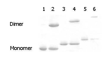
Figure 5 Western blot analysis of recombinant proteins CI, CII and CIII against anti-preS1 McAb (4D11) under reducing or nonreducing conditions.
Lanes 1, 3, and 5: purified CI, CII and CIII treated with 500 mmol/L DTT; lanes 2, 4, and 6: purified CI, CII and CIII not treated with DTT.
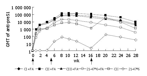
Figure 6 Dynamics of anti-preS1 responses of BALB/c mice immunized with CI, CII, CIII or 21-47*6 with Freund’s adjuvant (FA) at wk 0 and 5 (arrows) or immunized with CII and 21-47*6 without adjuvant at weeks 0, 5 and 16 (arrows).
Mice were immunized by i.m. injection of proteins: 20 μg CI, CII, CIII or 50 μg 21-47*6.
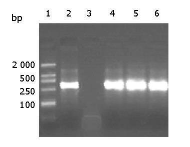
Figure 7 Detection of HBV captured by mouse antisera by PCR.
After immuno-capture of HBV from purified HBV virions, by antisera from three mice immunized with CII alone, HBV DNA was detected by PCR. 1: molecular markers 2: the positive control using anti-HBs monoclonal antibody 3: mouse serum injected with C149/mut 4-6: mouse sera immunized with CII alone at wk 2, 10 and 28 respectively.
- Citation: Yang HJ, Chen M, Cheng T, He SZ, Li SW, Guan BQ, Zhu Z-, Gu Y, Zhang J, Xia NS. Expression and immunoactivity of chimeric particulate antigens of receptor binding site-core antigen of hepatitis B virus. World J Gastroenterol 2005; 11(4): 492-497
- URL: https://www.wjgnet.com/1007-9327/full/v11/i4/492.htm
- DOI: https://dx.doi.org/10.3748/wjg.v11.i4.492


