Copyright
©The Author(s) 2005.
World J Gastroenterol. Oct 21, 2005; 11(39): 6090-6095
Published online Oct 21, 2005. doi: 10.3748/wjg.v11.i39.6090
Published online Oct 21, 2005. doi: 10.3748/wjg.v11.i39.6090
Figure 1 Endoscopic view of esophageal wall after MB and Lugol’s iodine double staining.
A: Before staining, a large broad-based erosion was observed on esophageal wall and the outlines of erosion were unclearly discernible; B: after MB staining, the same lesion stained more deeper blue than surrounding mucosa; C: after Lugol’s iodine double staining, the normal mucosa appeared brown, the same lesion stained more deeper blue than surrounding mucosa and the areas between both the colors were not stained.
Figure 2 Expression of GST-Π in histological esophageal tissues (?00).
A: Esophageal dysplasia showing yellow or brown granule in cell substance and nucleus; B: esophageal carcinoma showing yellow or brown granules in cell substance and nucleus.
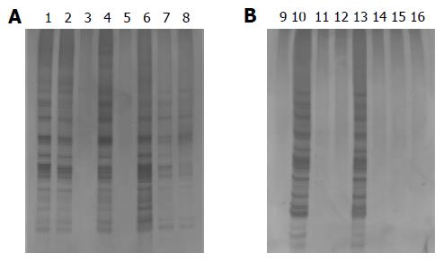
Figure 3 Analysis of telomerase activity of esophageal wall tissues using TRAP-silver staining assay.
A: Esophageal carcinoma showing telomerase positivity (lanes 1, 2, 4, 6-8) and telomerase negativity (lanes 3 and 5); B: esophageal dysplasia showing telomerase positivity (lanes 10 and 13), and telomerase negativity (lanes 9, 11, 12, 14, and 15), and negative control (lane 16).
- Citation: Zhu X, Zhang SH, Zhang KH, Li BM, Chen J. Value of endoscopic methylene blue and Lugol's iodine double staining and detection of GST-Π and telomerase in the early diagnosis of esophageal carcinoma. World J Gastroenterol 2005; 11(39): 6090-6095
- URL: https://www.wjgnet.com/1007-9327/full/v11/i39/6090.htm
- DOI: https://dx.doi.org/10.3748/wjg.v11.i39.6090


