
















©The Author(s) 2005.
World J Gastroenterol. Oct 21, 2005; 11(39): 6090-6095
Published online Oct 21, 2005. doi: 10.3748/wjg.v11.i39.6090
Published online Oct 21, 2005. doi: 10.3748/wjg.v11.i39.6090
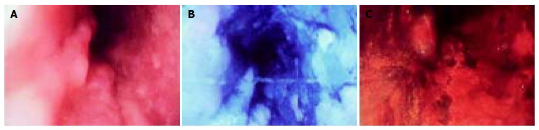
Figure 1 Endoscopic view of esophageal wall after MB and Lugol’s iodine double staining.
A: Before staining, a large broad-based erosion was observed on esophageal wall and the outlines of erosion were unclearly discernible; B: after MB staining, the same lesion stained more deeper blue than surrounding mucosa; C: after Lugol’s iodine double staining, the normal mucosa appeared brown, the same lesion stained more deeper blue than surrounding mucosa and the areas between both the colors were not stained.
- Citation: Zhu X, Zhang SH, Zhang KH, Li BM, Chen J. Value of endoscopic methylene blue and Lugol's iodine double staining and detection of GST-Π and telomerase in the early diagnosis of esophageal carcinoma. World J Gastroenterol 2005; 11(39): 6090-6095
- URL: https://www.wjgnet.com/1007-9327/full/v11/i39/6090.htm
- DOI: https://dx.doi.org/10.3748/wjg.v11.i39.6090