Copyright
©The Author(s) 2005.
World J Gastroenterol. Oct 7, 2005; 11(37): 5763-5769
Published online Oct 7, 2005. doi: 10.3748/wjg.v11.i37.5763
Published online Oct 7, 2005. doi: 10.3748/wjg.v11.i37.5763
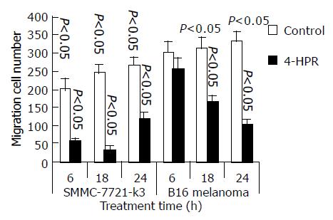
Figure 1 Inhibition migration of hepatocellular carcinoma and melanoma cells by 4-HPR treatment within a short period.
SMMC 7721-k3 and B16 melanoma cells were treated with 4-HPR for 6, 18, and 24 h, respectively. After treatment, the cells were assayed for migration in the Transwell.
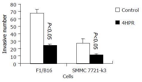
Figure 2 Effect of 3 mmol/L 4-HPR on cell invasion.
SMMC 7721-k3 and B16 melanoma cells were treated with 4-HPR for 6 h. The cells penetrating matrigel membrane and migrating to the bottom of the filter were counted and data were expressed as an average from six independent assays.
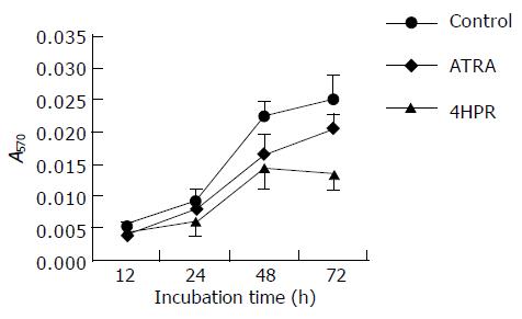
Figure 3 Growth inhibition of SMMC 7721-k3 cells by ATRA and 4-HPR.
SMMC 7721-k3 cells were cultured in the 96-well cluster plate and incubated with 10 mmol/L ATRA, 3 mmol/L 4-HPR, or ethanol in the control.
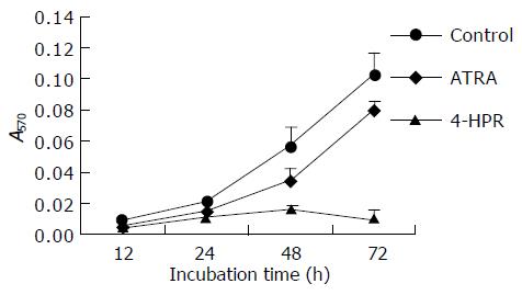
Figure 4 Growth inhibition of B16 melanoma cells by ATRA and 4-HPR.
B16 melanoma cells were cultured in the 96-well cluster plate and incubated with 10 mmol/L ATRA, 3 mmol/L 4-HPR, or ethanol in the control. The data were achieved by an average from eight independent groups.
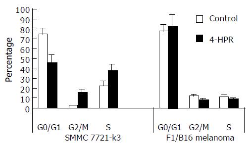
Figure 5 Regulation of cell cycles by 4-HPR.
SMMC 7721-k3 cells and B16 melanoma cells were treated with 4-HPR for 24 h, respectively. Data were expressed as an average from three independent assays.
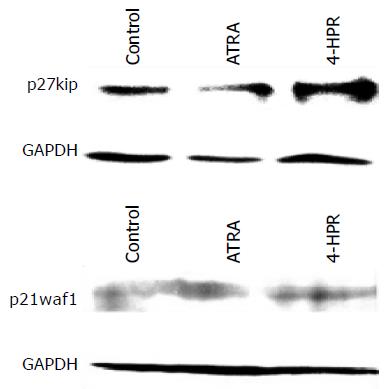
Figure 6 Western blot analysis of effects of 4-HPR on p27kip1 and p21waf1 expression.
SMMC 7721-k3 cells were treated with 10 and 3 mmol/L 4-HPR for 24 h, respectively.
Figure 7 Comparison of apoptosis peak between CST- and vector-transfected cells.
Control cells either of vector- or CST-transfected could not show any apoptosis (A). After being treated with 4-HPR, the vector-transfected cells produced a large apoptosis peak (B), while in CST-transfected cells only a small apoptosis peak could be seen (C).
-
Citation: Wu XZ, Zhang L, Shi BZ, Hu P. Inhibitory effects of
N -(4-hydrophenyl) retinamide on liver cancer and malignant melanoma cells. World J Gastroenterol 2005; 11(37): 5763-5769 - URL: https://www.wjgnet.com/1007-9327/full/v11/i37/5763.htm
- DOI: https://dx.doi.org/10.3748/wjg.v11.i37.5763


