Copyright
©The Author(s) 2005.
World J Gastroenterol. Sep 28, 2005; 11(36): 5638-5643
Published online Sep 28, 2005. doi: 10.3748/wjg.v11.i36.5638
Published online Sep 28, 2005. doi: 10.3748/wjg.v11.i36.5638
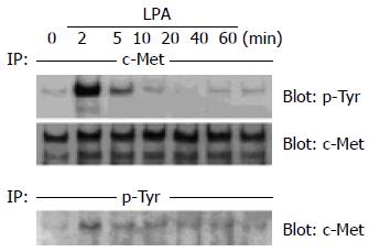
Figure 1 Rapid tyrosine phosphorylation of c-Met in response to LPA in human colon cancer cells.
(A) Human colon cancer LoVo cells were serum -starved for 24 h and then incubated with 10 µmol/L LPA for 2-60 min. After cell lysis, c -Met was immunoprecipitated (IP) using polyclonal anti-c -Met antibody, and immunoprecipitates were immunoblotted with monoclonal anti-phosphotyrosine antibody. Then the membrane was stripped and immunoblotted with anti-c-Met as a control. (B) Human colon cancer LoVo cells were serum-starved for 24 h and then incubated with 10 µmol/L LPA for 2-60 min. These cell lysates were IP with anti-phosphotyrosine, and the immunoprecipitates were immunoblotted with anti-c-Met.
Figure 2 Effect of PTX, a Gi inhibitor, on LPA-stimulated c-Met transactivation in colon cancer cells.
Human colon cancer LoVo cells were serum-starved for 24 h with or without 100 ng/mL PTX and then stimulated with 10 µmol/L LPA or 10 ng/mL HGF for 2 min. Cell lysates were IP with anti-c- Met. Immunoprecipitates were immunoblotted with anti-phosphotyrosine. Then the membrane was stripped and immunoblotted with anti-c -Met as a control. The same experiments were performed using anti-EGFR as a control for the inhibitory effect of PTX.
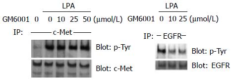
Figure 3 Effect of GM6001, an MMP inhibitor, on LPA-stimulated c-Met transactivation in colon cancer cells.
Human colon cancer LoVo cells were serum-starved for 24 h and pre-incubated with the indicated concentrations of GM6001 for 30 min. After incubation with 10 µmol/L LPA for 2 min, cell lysates were obtained and IP with anti-c-Met. Immunoprecipitates were immunoblotted with anti-phosphotyrosine. Then the membrane was stripped and immunoblotted with anti-c -Met as a control. The same experiments were performed using anti-EGFR as a control for the inhibitory effect of GM6001.
Figure 4 Effect of LPA on COX-2 expression in colon cancer cells.
Human colon cancer LoVo cells were serum-starved for 24 h, and then treated with 0.02-40 µmol/L LPA for 24 h. After cell lysis, the expression level of COX-2 protein was examined by Western blotting using anti-COX-2 antibody. Then the membrane was stripped and immunoblotted with anti-β-actin as a control.
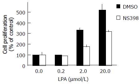
Figure 5 Inhibition of LPA-induced proliferation of LoVo cells by NS398, a COX-2 inhibitor.
Human colon cancer LoVo cells, after being pretreated with DMSO or NS398, were seeded with the indicated concentrations of LPA. After incubation for 120 h, the cell proliferation was measured using MTS assay. Data are expressed as a percentage of control, where control indicates cell proliferation in the absence of LPA without NS398 pretreatment (100%).
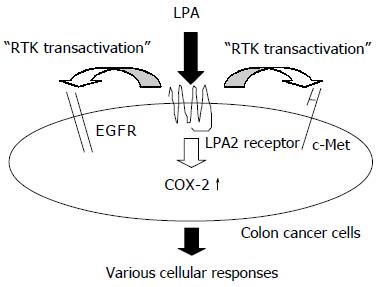
Figure 6 Mechanism of the effects of LPA on human colon cancer cells.
- Citation: Shida D, Kitayama J, Yamaguchi H, Yamashita H, Mori K, Watanabe T, Nagawa H. Lysophosphatidic acid transactivates both c-Met and epidermal growth factor receptor, and induces cyclooxygenase-2 expression in human colon cancer LoVo cells. World J Gastroenterol 2005; 11(36): 5638-5643
- URL: https://www.wjgnet.com/1007-9327/full/v11/i36/5638.htm
- DOI: https://dx.doi.org/10.3748/wjg.v11.i36.5638


