Copyright
©The Author(s) 2005.
World J Gastroenterol. Sep 28, 2005; 11(36): 5614-5620
Published online Sep 28, 2005. doi: 10.3748/wjg.v11.i36.5614
Published online Sep 28, 2005. doi: 10.3748/wjg.v11.i36.5614
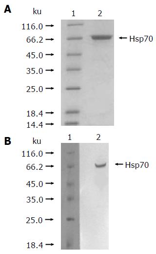
Figure 1 Purification of hsp70-peptide complexes from HCC cell line SMMC-7721.
(A) 12% reducing SDS-PAGE analysis of purified hsp70- peptide complexes preparation with silver staining. ( B) Western blotting analysis of purified hsp70-peptide complexes preparation. The preparation of hsp70-peptide complexes was immunoblotted with anti -hsp70 antibody. Lane 1 represents the protein molecular marker, and lane 2 represents hsp70-peptide complexes preparation.
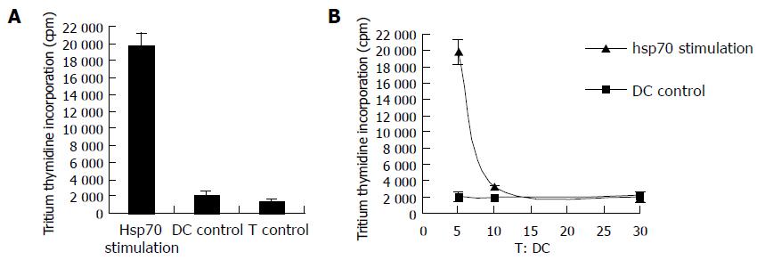
Figure 2 Stimulation of T cells by autologous DCs pulsed with hsp70-peptide complexes derived from SMMC-7721 cells.
DCs were incubated with 20 μg/mL hsp70-peptide complexes derived from SMMC- 7721 for 4 h at 37°C and cocultured with autologous T cells at different ratios for 5 d, and then tritiated thymidine was added and its incorporation was measured 18 h later. (A) 1 × 10⁵ T cells were cocultured with 2 × 10⁴ autologous DCs, pulsed with hsp70- peptide complexes (indicated as hsp70 stimulation) or unpulsed (indicated as DC control), or by themselves (indicated as T control) per well; (B) 1 × 10⁵ autologous T cells were cocultured with 2 × 10⁴, 1 × 10⁴, or 3.3 × 10³ DCs pulsed with hsp70-peptide complexes (indicated as hsp70 stimulation) or unpulsed (indicated as DC control) per well separately. The results are expressed as mean±SD and each value represents the mean of three replicates.
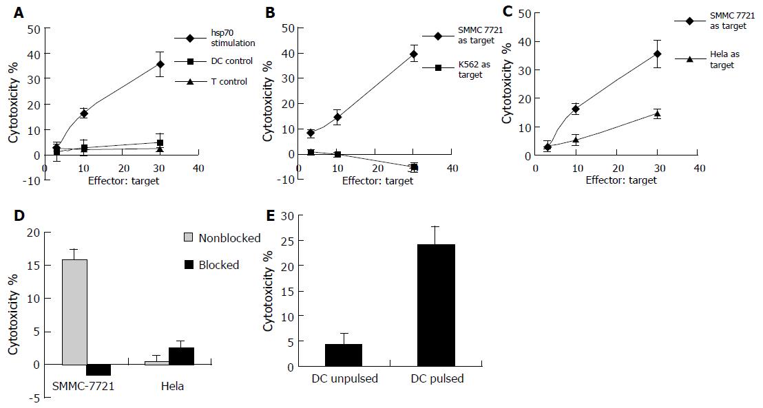
Figure 3 Generation of specific CTLs against SMMC-7721 by DCs pulsed with hsp70-peptide complexes derived from SMMC-7721 cells.
1 × 10⁵ DCs were incubated with 20 μg/mL hsp70- peptide complexes derived from SMMC-7721 for 4 h at 37°C and then cocultured with 1 × 10⁶ autologous T cells in each well of 24-well culture plate for 7-10 d in the presence of 20 U/mL human IL-2. The stimulated T cells were harvested by density gradient centrifugation and the cytotoxicity was determined by testing LDH release. As a control, T cells cocultured with unpulsed DCs or by themselves were also used as effectors. (A ) T cells were cocultured with autologous DCs pulsed with hsp70-peptide complexes (indicated as hsp70 stimulation) or unpulsed (indicated as DC control), or by themselves (indicated as T control), and SMMC -7721 cells were used as target cells; ( B) T cells were cocultured with autologous DCs pulsed with hsp70-peptide complexes, and SMMC-7721 cells (indicated as SMMC-7721 as target) or NK-sensitive cell line K562 cells (indicated as K562 as target) were used as target cells; (C) T cells were cocultured with autologous DCs pulsed with hsp70-peptide complexes and SMMC- 7721 cells (indicated as SMMC-7721 as target) or Hela cells (indicated as Hela as target) were used as target cells; (D) T cells cocultured with autologous DCs pulsed with hsp70-peptide complexes were used as effector cells and SMMC-7721 cells or Hela cells were used as target cells. The ratio of effector cells:target cells was 10:1. The target cells were preincubated with an anti-MHC class I antibody (W6/32; 1:50 dilution) or not, indicated as blocked and nonblocked separately. (E) T cells cocultured with autologous DCs pulsed with hsp70- peptide complexes were used as effector cells and autologous DCs pulsed with hsp70-peptide complexes or unpulsed were used as target cells (indicated as DC pulsed or DC unpulsed separately). The ratio of effector cells:target cells was 10:1. The results are expressed as mean±SD and each value represents the mean of three replicates.
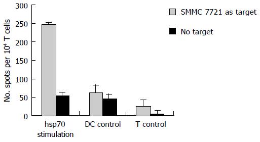
Figure 4 Secretion of IFN-γ by T cells induced with autologous DCs pulsed with hsp70-peptide complexes derived from SMMC-7721 cells.
1 × 10⁵ DCs were incubated with 20 μg/mL hsp70-peptide complexes derived from SMMC-7721 for 4 h at 37°C and then cocultured with 1 × 10⁶ autologous T cells in each well of 24-well culture plate for 7–10 d in the presence of 20 U/mL human IL-2. The stimulated T cells were harvested by density gradient centrifugation and their IFN-γ production was determined by ELISPOT assay (indicated as hsp70 stimulation). As a control, T cells cocultured with unpulsed DCs (indicated as DC control) or by themselves (indicated as T control) were also used as effectors. The results are expressed as mean±SD and each value represents the mean of three replicates.
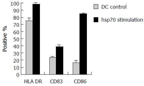
Figure 5 Maturation of human DCs by hsp70-peptide complexes derived from SMMC- 7721 cells.
DCs were incubated with 20 μg/mL hsp70- peptide complexes derived from SMMC-7721 cells for 4 h at 37°C, and cultured for 48 h. The DCs were then harvested and analyzed for expression of the indicated surface molecules using FACS analysis. The percentages shown were positive for the indicated surface markers. The results are expressed as mean±SD and each value represents the mean of three replicates.
- Citation: Wang XH, Qin Y, Hu MH, Xie Y. Dendritic cells pulsed with hsp70-peptide complexes derived from human hepatocellular carcinoma induce specific anti-tumor immune responses. World J Gastroenterol 2005; 11(36): 5614-5620
- URL: https://www.wjgnet.com/1007-9327/full/v11/i36/5614.htm
- DOI: https://dx.doi.org/10.3748/wjg.v11.i36.5614


