Copyright
©2005 Baishideng Publishing Group Inc.
World J Gastroenterol. Sep 21, 2005; 11(35): 5480-5484
Published online Sep 21, 2005. doi: 10.3748/wjg.v11.i35.5480
Published online Sep 21, 2005. doi: 10.3748/wjg.v11.i35.5480
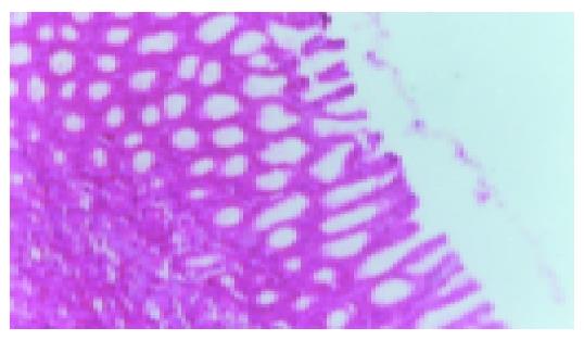
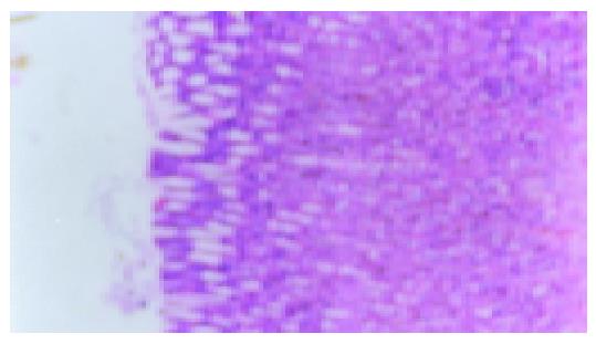
Figure 1 Normal gastric mucous (×200).
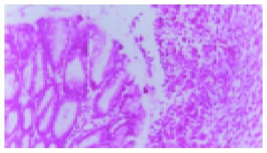
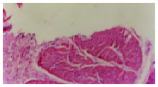
Figure 2 Model group ulcer formation (7 d, ×200).
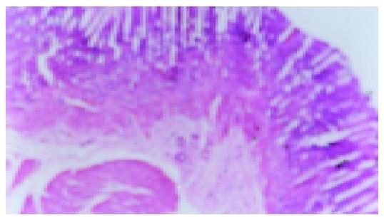
Figure 3 Model group without IL-b (92 d), scar tissue under the thinned gastric mucosa (×200).
Figure 4 Ranitidine group with IL-b, small ulcer can be seen (×200).
Figure 5 JWYY group, after ip IL-b, no ulcer recurrence (×200).
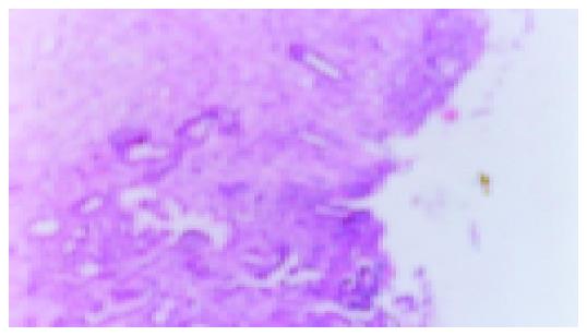
Figure 6 Model recurrence group, after ip IL-b, ulcer relapses (×200).
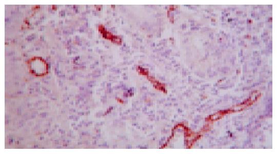
Figure 7 Microvessel of gastric ulcer margin in the rat stained by VIII factor (method of SABC ×400).
- Citation: Dai XP, Li JB, Liu ZQ, Ding X, Huang CH, Zhou B. Effect of Jianweiyuyang granule on gastric ulcer recurrence and expression of VEGF mRNA in the healing process of gastric ulcer in rats. World J Gastroenterol 2005; 11(35): 5480-5484
- URL: https://www.wjgnet.com/1007-9327/full/v11/i35/5480.htm
- DOI: https://dx.doi.org/10.3748/wjg.v11.i35.5480


