Copyright
©The Author(s) 2005.
World J Gastroenterol. Aug 21, 2005; 11(31): 4927-4929
Published online Aug 21, 2005. doi: 10.3748/wjg.v11.i31.4927
Published online Aug 21, 2005. doi: 10.3748/wjg.v11.i31.4927
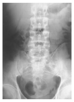
Figure 1 A round mass in pelvis with rim calcification on the abdominal plain radiograph.
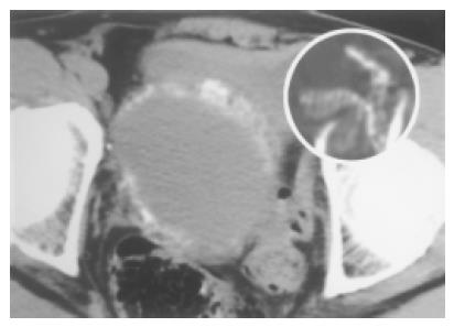
Figure 2 A cystic lesion in pelvic cavity with a thick “calcified reticulate rind” demonstrated by pre-enhanced CT scan.
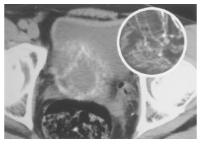
Figure 3 “Calcified reticulate rind” on another level (2 cm below Figure 2) demonstrated by pre-enhanced CT scan.
- Citation: Lu YY, Cheung YC, Ko SF, Ng SH. Calcified reticulate rind sign: A characteristic feature of gossypiboma on computed tomography. World J Gastroenterol 2005; 11(31): 4927-4929
- URL: https://www.wjgnet.com/1007-9327/full/v11/i31/4927.htm
- DOI: https://dx.doi.org/10.3748/wjg.v11.i31.4927


