Copyright
©The Author(s) 2005.
World J Gastroenterol. Aug 21, 2005; 11(31): 4827-4832
Published online Aug 21, 2005. doi: 10.3748/wjg.v11.i31.4827
Published online Aug 21, 2005. doi: 10.3748/wjg.v11.i31.4827
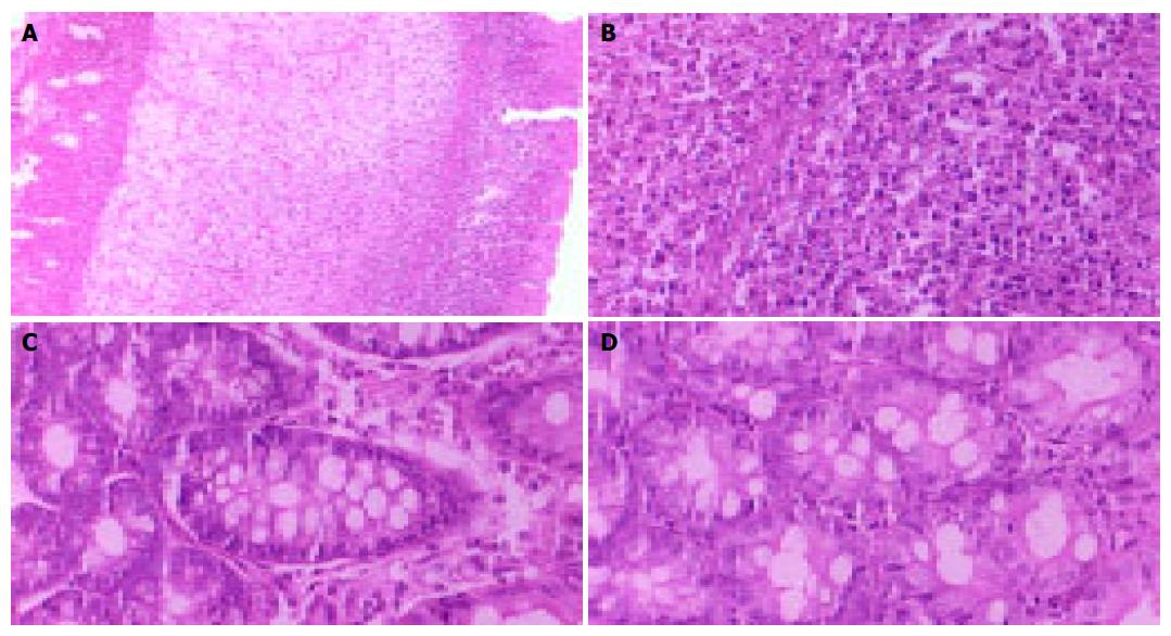
Figure 1 Histopathological changes of the colon.
A: After 3 d of TNBS administration, the structure of the colon was destroyed (×10); B: Numerous inflammatory cells were infiltrated in the submucosa and mucosa of the colon (×40); C: After 28 d of TNBS administration, only a few inflammatory cells were infiltrated in submucosa and mucosa of the colon (×40); D: The inflammatory cells were minimal in the colon of the rats after 3 d of saline administration (×40).
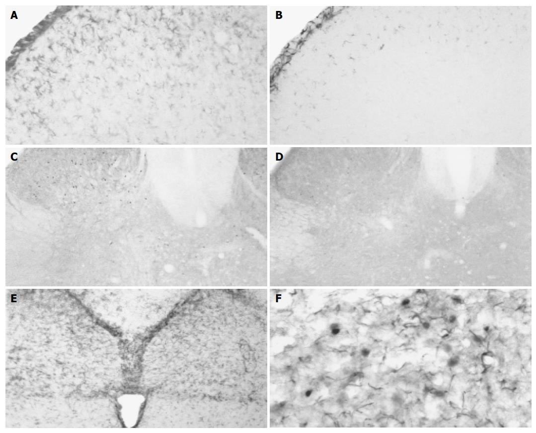
Figure 2 GFAP and Fos expression in the spinal cord and NTS.
A: After 3 d of TNBS administration, most activated astrocytes positive for GFAP were distributed in the superficial laminae (laminae I-II) of dorsal horn (×20); B: Mild GFAP-IR astrocytes exhibited in the spinal cord of rats after 3 d of saline administration (×20); C: After 14 d of TNBS administration, Fos-IR cells were mainly localized in deeper laminae III-IV and laminae V-VI in the spinal dorsal horn (×10); D: Only a few Fos-IR neurons were sparsely distributed in the dorsal horn after 7 d of saline administration (×10); E: After 3 d of TNBS administration, GFAP-IR glial cells increased and were primarily localized in NTS (×10); F: After 3 d of TNBS administration, activated astrocytes positive for GFAP enveloped activated neurons stained for Fos in the medulla oblongata (×40).
- Citation: Sun YN, Luo JY, Rao ZR, Lan L, Duan L. GFAP and Fos immunoreactivity in lumbo-sacral spinal cord and medulla oblongata after chronic colonic inflammation in rats. World J Gastroenterol 2005; 11(31): 4827-4832
- URL: https://www.wjgnet.com/1007-9327/full/v11/i31/4827.htm
- DOI: https://dx.doi.org/10.3748/wjg.v11.i31.4827


