Copyright
©The Author(s) 2005.
World J Gastroenterol. Aug 7, 2005; 11(29): 4524-4529
Published online Aug 7, 2005. doi: 10.3748/wjg.v11.i29.4524
Published online Aug 7, 2005. doi: 10.3748/wjg.v11.i29.4524
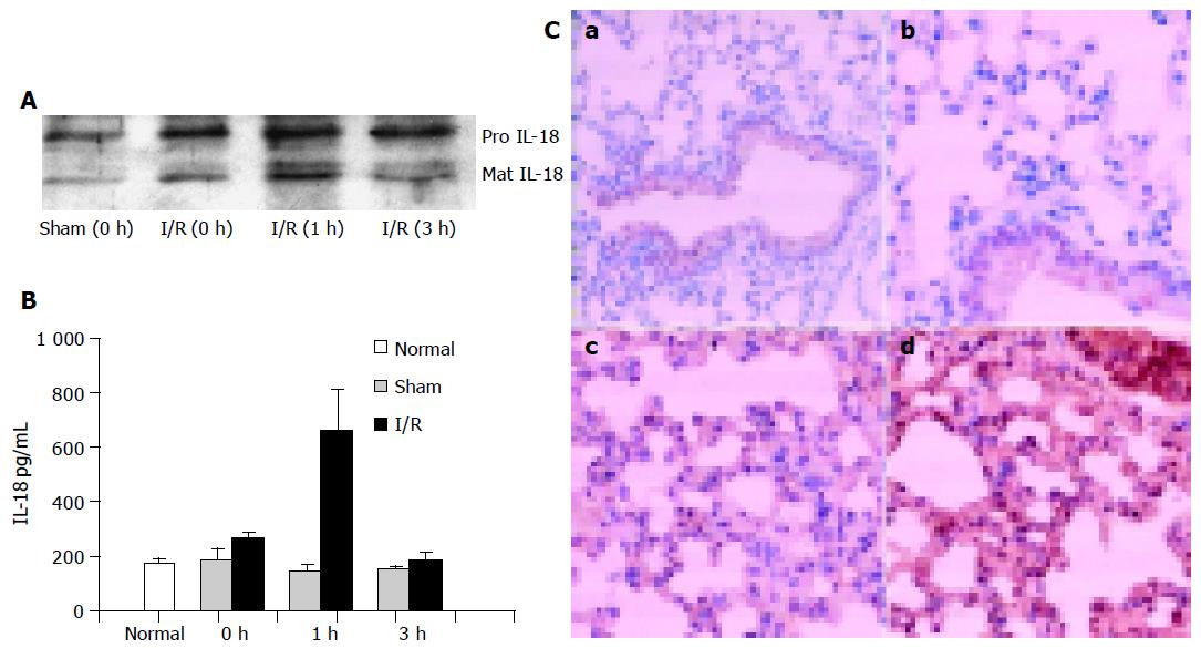
Figure 1 Gut ischemia/reperfusion results in increased levels of IL-18 in serum and lung.
(A and B) The circulating levels of IL-18 after gut ischemia/reperfusion. Mice were subjected to gut ischemia/reperfusion or sham. Serum IL-18 at the indicated times after reperfusion was collected, part of which was immunoprecipitated with polyclonal rabbit anti-murine IL-18 antibody and analyzed by immunoblotting (A), and another part was assayed with ELISA (B) (n = 5). (C) Gut ischemia/reperfusion results in elevated levels of IL-18 in lung tissue. Immunohistochemistry of lung sections obtained from normal (a, ×100, and b, ×200), 1 h (c) or 3 h (d) I/R model. The positive staining for IL-18 shows as dark brown. Magnification (c and d) ×200.
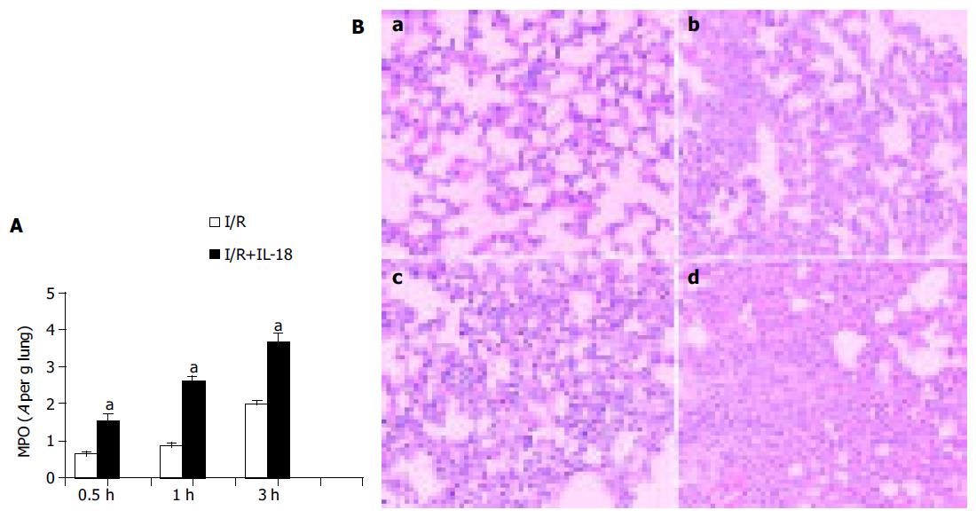
Figure 2 The effect of exogenous IL-18 on the ischemia/reperfusion-induced lung injury.
A: IL-18 injection further remarkably enhanced the neutrophil sequestration in lung. After indicated times of reperfusion, the MPO activity was determined in lung tissue (n = 5), aP<0.05 vs I/R. B: Comparison of pulmonary histopathology. Lungs from IL-18-nontreated (a, for 1 h of reperfusion and b, for 3 h) or IL-18-treated (c, for 1 h and d, for 3 h of reperfusion) mice subjected to gut I/R and stained with HE. Magnification ×100.
Figure 3 The effect of anti-IL-18 antibody on the ischemia-induced lung injury.
A: Anti-IL-18 antibody injection remarkably inhibited the lung MPO activity. After 3 h of reperfusion, the MPO activity was determined in lung tissues (n = 5), aP<0.05 vs others. B: Comparison of pulmonary histopathology. Lungs from ischemic mice (a, for 3 h of reperfusion), and ischemic mice treated with IL-18Ab (b, for 3 h of reperfusion) and stained with HE. Magnification ×100.
- Citation: Yang YJ, Shen Y, Chen SH, Ge XR. Role of interleukin 18 in acute lung inflammation induced by gut ischemia reperfusion. World J Gastroenterol 2005; 11(29): 4524-4529
- URL: https://www.wjgnet.com/1007-9327/full/v11/i29/4524.htm
- DOI: https://dx.doi.org/10.3748/wjg.v11.i29.4524


