Copyright
©The Author(s) 2005.
World J Gastroenterol. Aug 7, 2005; 11(29): 4457-4460
Published online Aug 7, 2005. doi: 10.3748/wjg.v11.i29.4457
Published online Aug 7, 2005. doi: 10.3748/wjg.v11.i29.4457
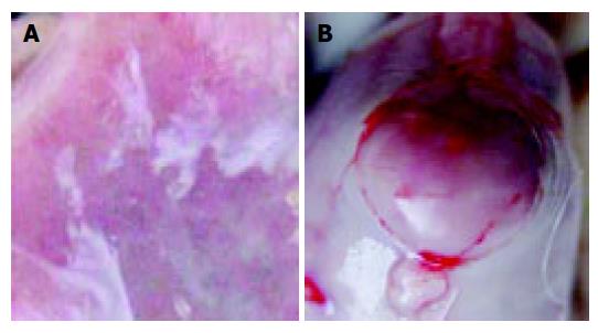
Figure 1 Erosive mucosa of rat stomach (A) and leiomyoma under serosa (B).
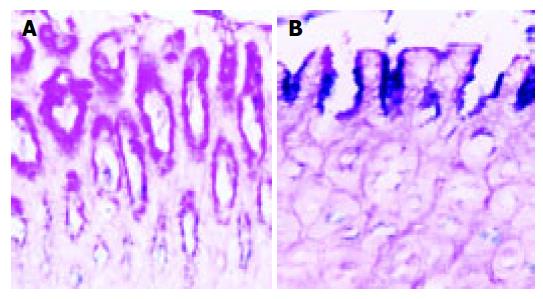
Figure 2 Normal mucosa (A) and erosive mucosa (B) of rat stomach, AB-PAS, ×200.
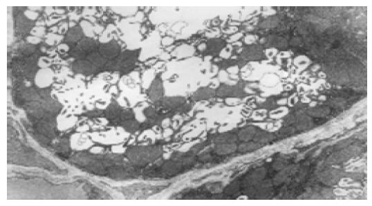
Figure 3 Intracellular secretory canaliculi of parietal cells in normal control group, TEM 5.
8×10³.
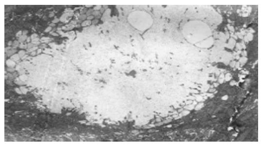
Figure 4 Expansion of intracellular secretory canaliculi of parietal cells in high selenium group, TEM 5.
8×10³.
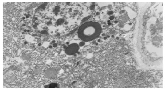
Figure 5 Endocrine cells and secondary lysosomes in high selenium group, TEM 10×103.
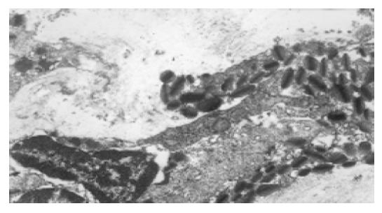
Figure 6 Infiltration of eosinophils in gastric mucosa of high selenium group, TEM 10×103.
- Citation: Su YP, Tang JM, Tang Y, Gao HY. Histological and ultrastructural changes induced by selenium in early experimental gastric carcinogenesis. World J Gastroenterol 2005; 11(29): 4457-4460
- URL: https://www.wjgnet.com/1007-9327/full/v11/i29/4457.htm
- DOI: https://dx.doi.org/10.3748/wjg.v11.i29.4457


