Copyright
©The Author(s) 2005.
World J Gastroenterol. Jul 14, 2005; 11(26): 4098-4101
Published online Jul 14, 2005. doi: 10.3748/wjg.v11.i26.4098
Published online Jul 14, 2005. doi: 10.3748/wjg.v11.i26.4098
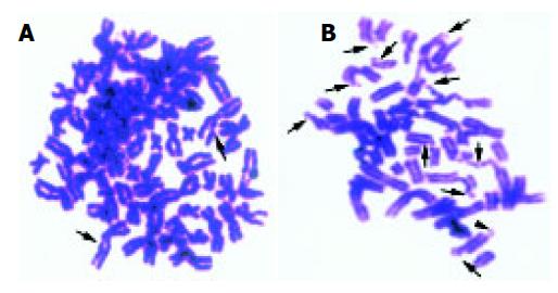
Figure 1 G2 phase prematurely condensed chromosome spreads.
A: SMMC-7721 cells after exposed to the γ-rays of 0.5 Gy; B: L02 cells after exposed to the γ-rays of 8 Gy. Arrows show the chromatid breaks, arrowheads show the isochromatid breaks.
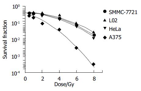
Figure 2 Cell survival curve after exposed to γ-rays.
All of four cell lines have a linear quadratic survival curve, which were good agreement with classical model.
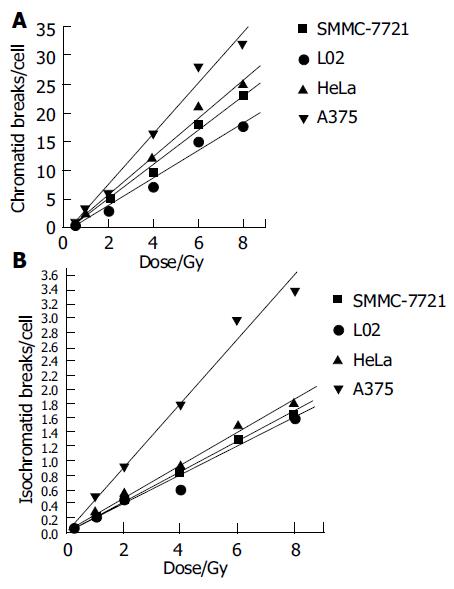
Figure 3 G2 chromosome breaks at various absorbed dose.
A: chromatid breaks; B: isochromatid breaks.
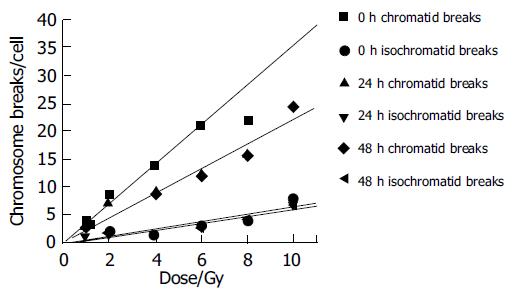
Figure 4 Correlation between chromatid breaks and absorbed dose at 0, 24, and 48 h after exposed to radiation.
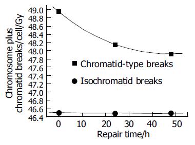
Figure 5 Kinetic repair of chromosome breakages within 48 h after L02 cell line was exposed to γ-rays.
The chromatid breaks plus 46 (number of normal human chromosomes) make this change remarkably.
- Citation: Yang JS, Li WJ, Zhou GM, Jin XD, Xia JG, Wang JF, Wang ZZ, Guo CL, Gao QX. Comparative study on radiosensitivity of various tumor cells and human normal liver cells. World J Gastroenterol 2005; 11(26): 4098-4101
- URL: https://www.wjgnet.com/1007-9327/full/v11/i26/4098.htm
- DOI: https://dx.doi.org/10.3748/wjg.v11.i26.4098


