Copyright
©2005 Baishideng Publishing Group Inc.
World J Gastroenterol. Jun 28, 2005; 11(24): 3800-3802
Published online Jun 28, 2005. doi: 10.3748/wjg.v11.i24.3800
Published online Jun 28, 2005. doi: 10.3748/wjg.v11.i24.3800
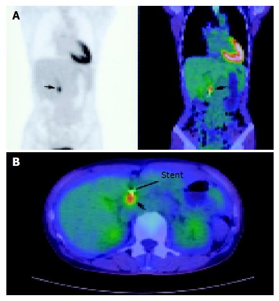
Figure 1 Whole body and combined fusion CT-PET of the pancreas.
A: Whole body CT-PET demonstrating a focus of increased uptake in the pancreatic head (arrowed); B: Although no lesion was seen on CT, anatomical localization was possible with fusion CT-PET to the pancreatic head (arrowed). A CBD stent is seen in situ.
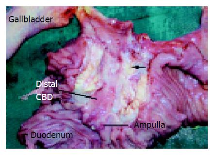
Figure 2 Post-Whipple’s specimen demonstrating a mass in the head of pancreas causing narrowing of the distal CBD (arrowed).
- Citation: Goh BKP, Tan YM, Chung YFA. Utility of fusion CT-PET in the diagnosis of small pancreatic carcinoma. World J Gastroenterol 2005; 11(24): 3800-3802
- URL: https://www.wjgnet.com/1007-9327/full/v11/i24/3800.htm
- DOI: https://dx.doi.org/10.3748/wjg.v11.i24.3800


