Copyright
©2005 Baishideng Publishing Group Inc.
World J Gastroenterol. Jun 14, 2005; 11(22): 3446-3450
Published online Jun 14, 2005. doi: 10.3748/wjg.v11.i22.3446
Published online Jun 14, 2005. doi: 10.3748/wjg.v11.i22.3446
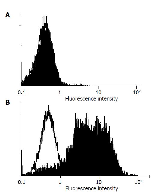
Figure 1 FasL expression on the surface of adenovirus infected SGC-7901 cells.
A: Ad-LacZ (titer 4.0×10⁵ CFU/mL); B: Ad-FasL (titer 2.8×10⁵ CFU/mL).
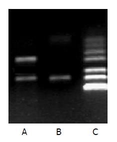
Figure 2 FasL expression as examined by RT-PCR.
A: SGC-7901-Fas-L (the FasL amplicon has a size of 231 bp); B: SGC-7901-vect (negative control); C: DNA marker.
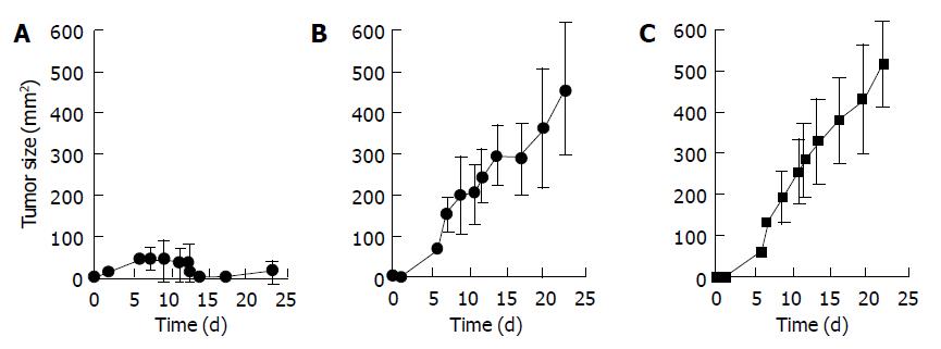
Figure 3 Treatment of SGC-7901 tumor-bearing mice with SGC-7901+FasL cells.
A: Mice were injected with SGC-7901 cells (5×10⁵), followed by treatment with FasL (n = 8); B: mice were injected with SGC-7901 cells (5×10⁵); C: mice were injected with SGC-7901 cells (5×10⁵), followed by treatment with PBS (n = 8). Tumor incidence is presented at each time point in Figure. P<0.0001.
- Citation: Zheng SY, Li DC, Zhang ZD, Zhao J, Ge JF. Adenovirus-mediated FasL gene transfer into human gastric carcinoma. World J Gastroenterol 2005; 11(22): 3446-3450
- URL: https://www.wjgnet.com/1007-9327/full/v11/i22/3446.htm
- DOI: https://dx.doi.org/10.3748/wjg.v11.i22.3446


