Copyright
©2005 Baishideng Publishing Group Inc.
World J Gastroenterol. Jun 14, 2005; 11(22): 3363-3367
Published online Jun 14, 2005. doi: 10.3748/wjg.v11.i22.3363
Published online Jun 14, 2005. doi: 10.3748/wjg.v11.i22.3363
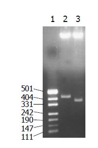
Figure 1 Restriction analysis of recombinant plasmids pET-RNA-I1.
Lane 1: DNA molecular weight markers pUC19 DNA/Msp I (Hpa ll); lane 2: pET-RNA-I1-E1 digested with SpeI/MluI (410 bp); lane 3: pET-RNA-I1- wt digested with SpeI/MluI (365 bp).
Figure 2 Sequence analysis of pET-RNA-I1-E1.
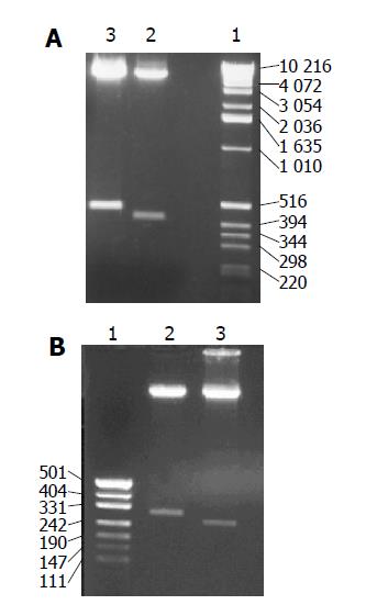
Figure 3 Restriction analysis of recombinant plasmid pET-RNA-I2 (A) and pET-RNA-I3 (B).
A: Restriction analysis of recombinant plasmid pET-RNA-I2. Lane 1: DNA molecular weight markers (1 kb); lane 2: pET-RNA-I2- wt digested with SpeI/ NcoI (427bp); lane 3: pET-RNA-I2-E1 digested with SpeI/NcoI (472bp); B: Restriction analysis of recombinant plasmid pET-RNA-I3. Lane 1: DNA molecular weight markers pUC19 DNA/MspI (Hpall); lane 2: pET-RNA-I3-E1 digested with NsiI/NcoI (302 bp); lane 3: pET-RNA-I3- wt digested with NsiI/NcoI (257 bp).
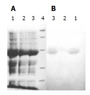
Figure 4 SDS-PAGE of inclusion body of RNA-E1 (A) and Western blot of chimeric antigen protein RNA-E1(B).
A: SDS-PAGE of inclusion body of RNA-E1. Lane 1: Inclusion bodies of RNA-I1-E1; lane 2: Inclusion bodies of RNA-I2-E1; lane 3: Inclusion bodies of RNA-I3-E1; lane 4: M: Protein molecular weight standard (middle range); B: Western blot of chimeric antigen protein RNA-E1 Lane 1: RNA-I1-E1; lane 2: RNA-I2-E1; lane 3: RNA-I3-E1.
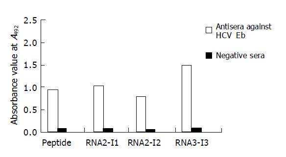
Figure 5 ELISA of E1 peptide and RNA-E1 chimeric antigen displaying epitope at three different positions.
- Citation: Peng M, Dai CB, Chen YD. Expression and immunoreactivity of an epitope of HCV in a foreign epitope presenting system. World J Gastroenterol 2005; 11(22): 3363-3367
- URL: https://www.wjgnet.com/1007-9327/full/v11/i22/3363.htm
- DOI: https://dx.doi.org/10.3748/wjg.v11.i22.3363


